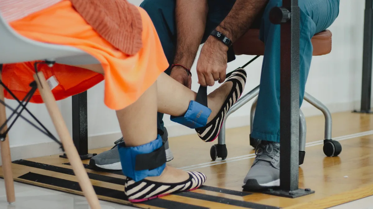
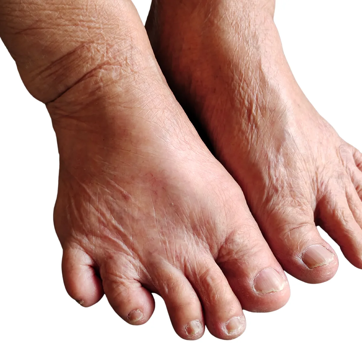
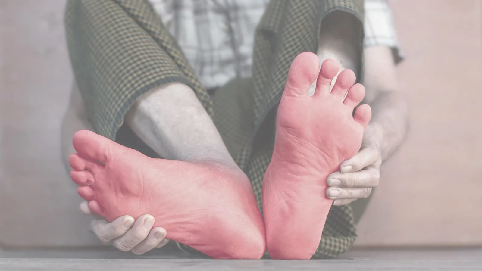
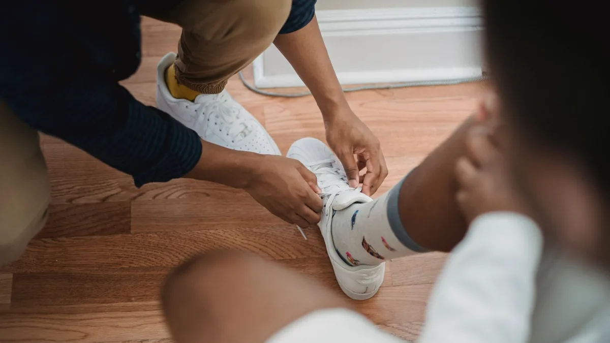
When peripheral neuropathy dulls protective feeling, footwear stops being a comfort choice and becomes injury prevention. The shoe you want holds the heel still and leaves room at the toes. It spreads pressure out instead of focusing it, and it never needs breaking in. Pair that shoe with a look at both feet every single day, because a numb foot no longer reports its own injuries.
Knowing this is not the same as doing it. A cross-sectional study looked at 212 patients with diabetes in a row at a rural hospital in Tamil Nadu, southern India. Of them, 75% scored well on foot-care knowledge. Yet only 67% scored well on foot-care practice. A good score meant at least half the possible points [1].
Key takeaways
Dulled protective feeling changes how rubbing, pressure, and heat register. So skin injury can start before you feel anything.
Foot care starts with finding the cause behind your neuropathy, because neuropathy is a category, not a single diagnosis.
Daily checks are the safety system that stands in for the warning your nerves can no longer give.
Fit matters more than brand: room at the toes, a heel that does not slip, and closure you can control.
Electrodiagnostic testing helps show which nerve fibers are involved. That shapes how hard the foot needs to be protected.
Burning feet at night and dulled feeling can exist together. So comfort at rest is not proof that the skin is safe during the day.
Why do shoes matter so much with peripheral neuropathy?
Footwear is part of the mechanical side of neuropathy care. The nerve fibers that report pressure, heat, and rubbing are exactly the ones peripheral neuropathy tends to hit first in the feet.
When protective feeling drops, the foot keeps taking small injuries long after they would once have hurt. That is the silent injury path. It is why a shoe problem can become a skin problem without a single painful moment.
Three ways the wrong shoe injures a numb foot
Friction and shear cause small skin injuries that never register as pain.
Focal pressure piles stress on one spot, especially where there is a bunion, a hammertoe, or a thickened nail.
Heat and trapped moisture soften and break down skin. They can also make burning feel worse in the evening.
What your symptom pattern changes in real life
Peripheral neuropathy of the feet often hits the longest nerves first, so the toes go first. Small-fiber symptoms tend to be burning, electric, or hot-coal feelings. Large-fiber loss shows up as numbness and dulled protective feeling.
Those two patterns call for different focus. If you cannot reliably feel injury, prevention has to come before comfort. For how these symptoms show up and what they suggest, see peripheral neuropathy of the feet symptoms.
How should shoes fit when you have neuropathy?
Good footwear for a numb foot is a no-surprises zone. It is stable, supportive, and predictable from the first minute you wear it. Fit does more of that work than any material or brand claim.
Fit checks to run before you wear a shoe
Toe box space: your toes should not feel compressed at any point in the stride.
Heel stability: the heel should not lift or slide, because sliding is what causes shear.
Closure control: laces or straps should let you keep pressure even across the top of the foot.
No break-in period: do not break in shoes on numb feet. If something rubs, stop wearing them and look at the skin.
What to avoid when sensation is reduced
Loose sandals that let the foot slide side to side.
Hard, narrow shoes that concentrate pressure on a deformity.
Soft slippers with no structure, which let the foot shift and rub.
Any shoe that feels fine at first and uncomfortable later, since a delayed or muted signal is exactly what neuropathy causes.
Daily foot inspection is your safety system
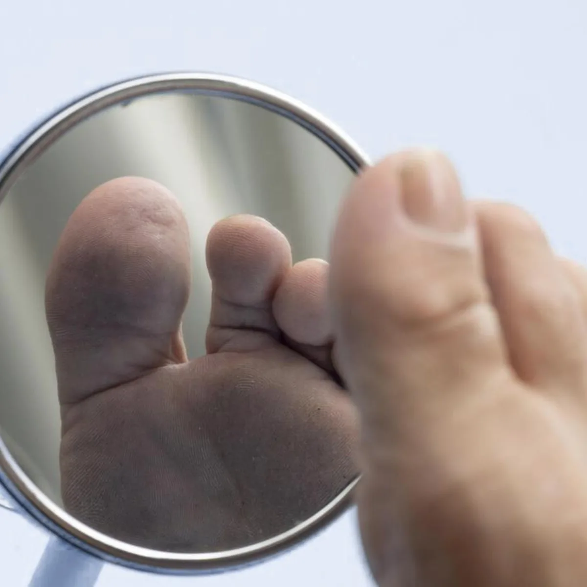
Check both feet at the same time every day, including between the toes and across the sole. If you cannot see the sole, use a mirror and a bright light, or ask someone to look for you.
Checking matters most for the people who feel the least. It also matters when burning at night is the loudest symptom, because an injury picked up in the morning may give no warning at all.
What causes peripheral neuropathy in the feet?
Neuropathy is a category, not a diagnosis. Two people can have the same numbness in the toes and totally different problems behind it. That is why the workup looks for the cause first.
The feet go first when the longest nerves are hit, so they are where an untreated cause shows up soonest. That is the case for mapping the cause instead of only managing the feeling.
Drivers that change footwear and foot-care priorities
Metabolic and diabetic drivers, where poor tiny-vessel blood supply and metabolic stress affect the tissue around the nerve.
Nutritional drivers, including B12-related patterns that can add to nerve problems.
Gluten-related nerve injury, which can matter even when celiac testing is negative.
Toxic and medication-related exposures, including alcohol-related nerve injury and chemotherapy history.
Autoimmune and structural contributors, which can cause patterns that behave differently in the foot.
Small-fiber predominant patterns, which tend toward burning. Large-fiber loss, by contrast, raises the risk of a wound you do not notice.
Diabetic peripheral neuropathy involves both metabolic and tiny-vessel stress. The feet are simply where it shows. When less oxygen reaches the nerve and skin, healing after a small injury is slower and less reliable.
What this means for footwear choices
Choose stability: a stable shoe cuts shear during walking, which is the injury most often missed.
Inspect earlier: look for redness, blisters, and callus even when nothing hurts.
Manage moisture: dry skin cracks and damp skin gets soggy, and both increase rubbing damage.
Plan around deformity: thickened nails and bunions shift pressure and may call for a modified shoe or insert.
When should you see a doctor about a neuropathic foot problem?
An open sore, or redness that is spreading.
New swelling, especially with a change in skin temperature.
Blisters that keep returning in the same spot in the same shoe.
A sudden change in numbness or feeling after a change in footwear.
Which tests help plan foot protection?
Foot protection gets more precise once the fiber type and the cause are clearer. Vague labels lead to vague plans.
Electrodiagnostic testing and why the pattern drives the plan
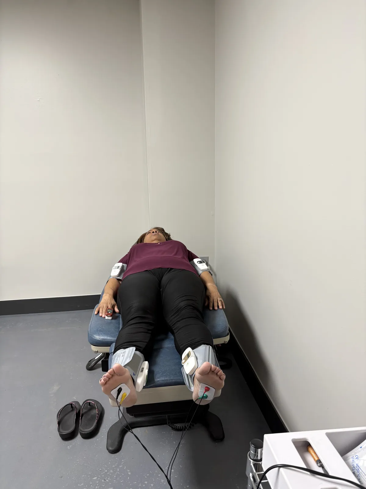
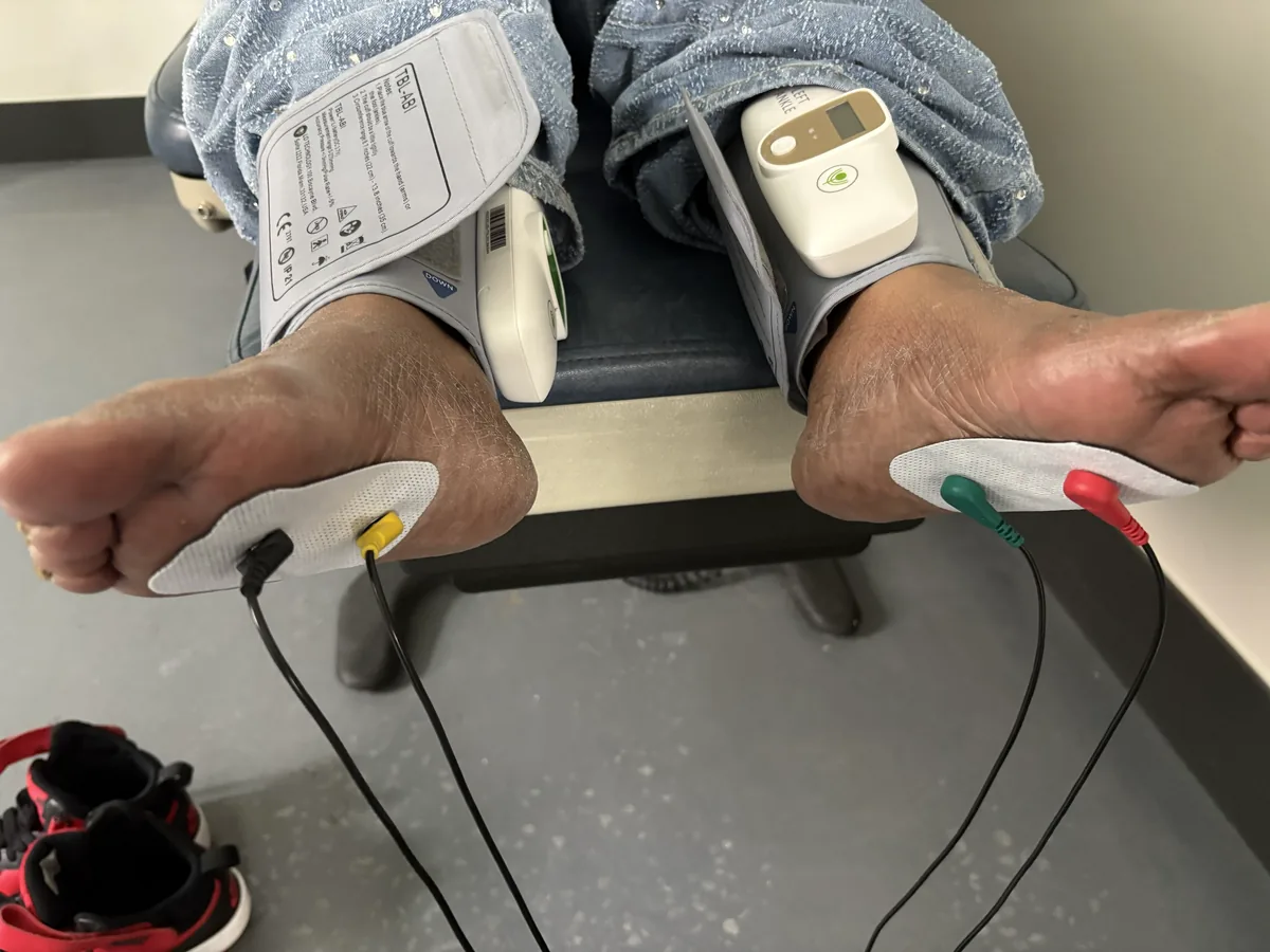
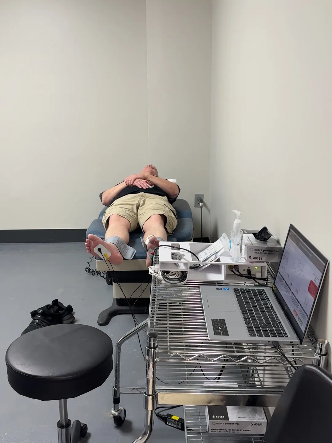
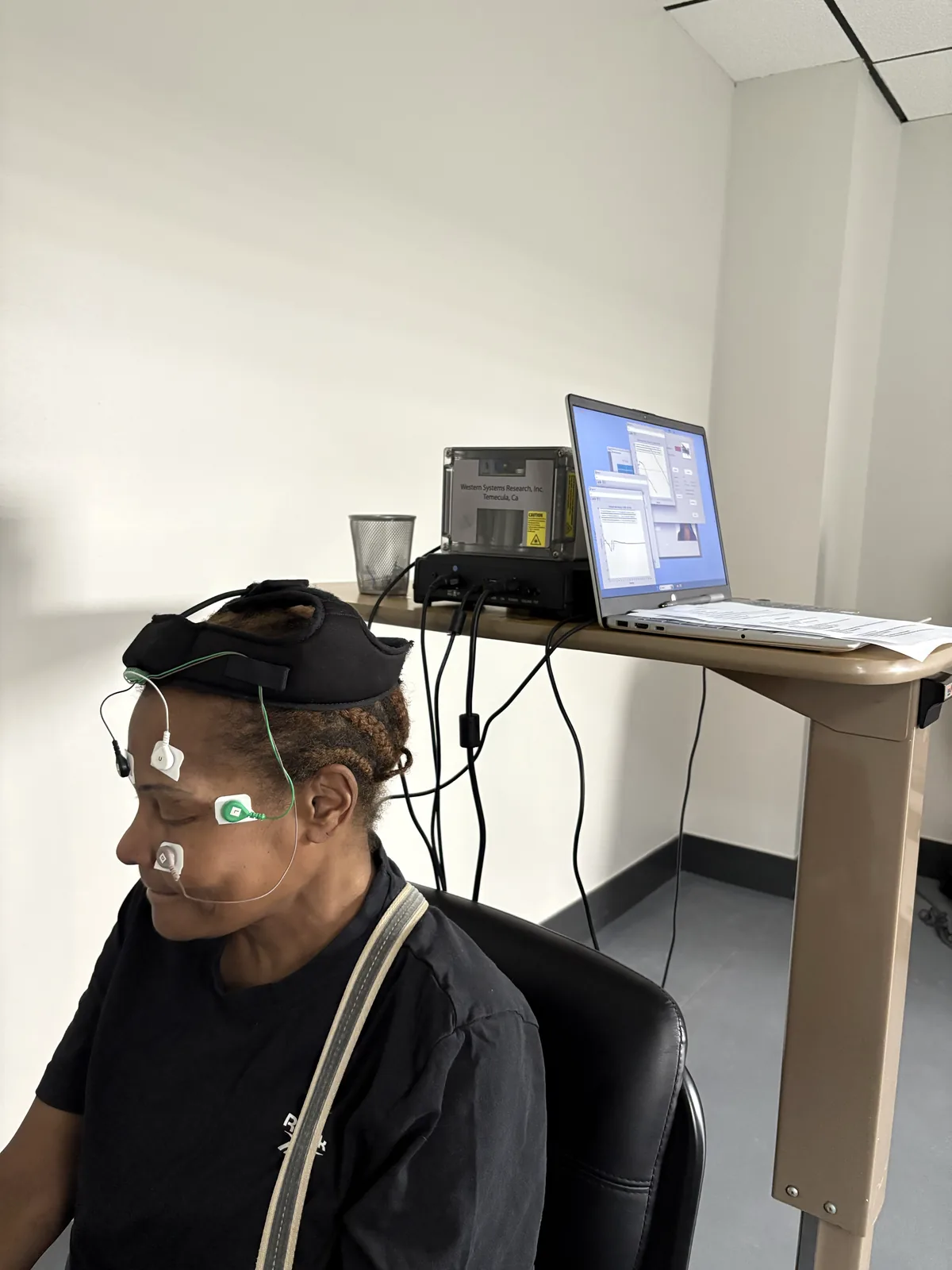
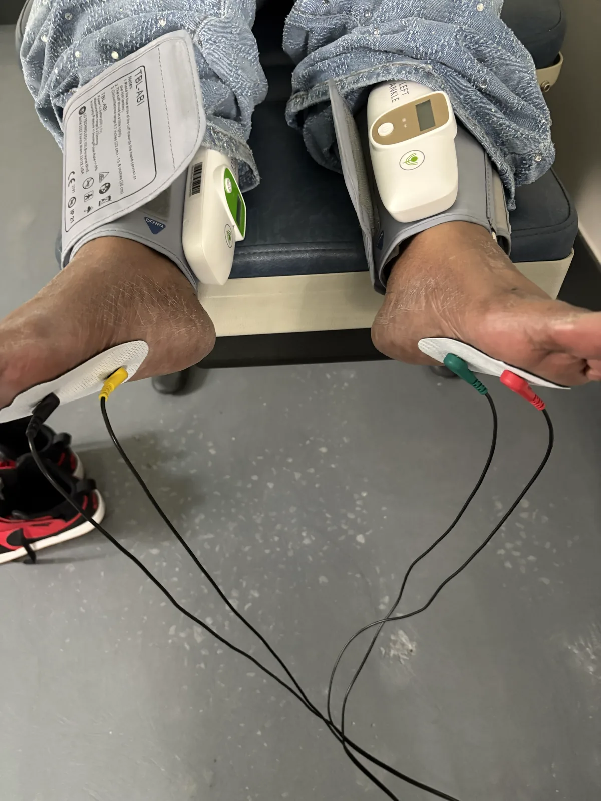
Electrodiagnostic testing means EMG and nerve conduction studies. We do it on site at Regenerve. It gives information about large-fiber damage. Small-fiber damage is not always picked up the same way, so we choose the tests to match the question being asked.
How in-clinic therapies connect to foot protection
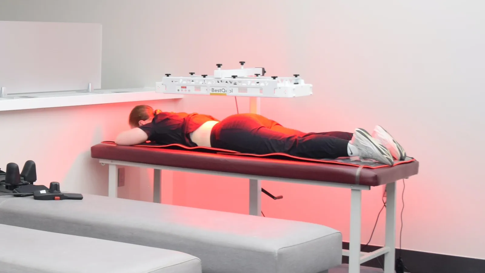
Regenerve uses class 4 photobiomodulation, class 3B cold laser, whole-body infrared, and metabolic and nutrition care as the medical side of the plan. None of these replaces a well-fitted shoe or a daily check.
The two halves do different jobs. The medical plan works on the environment around the nerve. The shoe and the mirror work on the physical harm that happens between visits.
Putting it together in the St. Louis region
Three things run side by side: protect the foot physically, find the cause medically, and check your feet every day. Dropping any one of them tends to undo the other two.
How a foot protection plan gets built
Screen for the most likely main cause with the free five-question Nerve Damage Score, then review the result together.
Clarify the nerve fiber pattern with testing that fits your symptoms.
Build the footwear and foot-check plan around your level of feeling, any deformities, and your risk factors.
Reassess and adjust, because both the cause and the foot can change over time.
Frequently asked questions
What kind of shoes help when you have peripheral neuropathy?
Look for stable support, a roomy toe box, and a closure that stops the foot from sliding, since rubbing and focused pressure are what damage skin that cannot report pain. Fit is checked on your foot, not on the box. For how footwear fits with the rest of the plan, see peripheral neuropathy treatments for the feet.
Should numbness be treated with medication or with footwear first?
Both have roles, but physical protection comes first, because medicine does not restore the warning signal that prevents a wound. Numb feet need a shoe and a daily check before they need anything else. For what numbness and burning each suggest, see small fiber neuropathy symptoms and testing.
Is walking safe when my feet are numb?
Usually yes, in well-fitted shoes and with both feet checked before and after. Any open wound, or a hot and swollen foot, needs a checkup before weight-bearing activity. For a step-by-step approach to movement, see exercises for peripheral neuropathy in the feet.
What should I do if my feet burn at night but the skin looks normal?
Burning at night is common in small-fiber patterns and does not mean an injury will be visible. So daily checks and friction control still apply. It is also a reason to look for the cause instead of settling for symptom control. For what a workup includes, see the services offered at Regenerve.
Do shoes for neuropathy really work?
Shoes do a physical job. They cut down the rubbing, shear and focused pressure that injure skin you can no longer feel. Fit matters more than brand. Look for room at the toes, a heel that does not slip and a closure you control. A shoe does not treat the nerve, which is why the medical plan and the shoe work side by side.
What are the worst shoes for people with neuropathy?
Loose sandals that let the foot slide, hard narrow shoes that press on a bunion or hammertoe, and soft slippers with no structure all allow rubbing and pressure you may not feel. So does any shoe that needs breaking in, or one that feels fine at first and uncomfortable later. If a shoe rubs, stop wearing it and check the skin.
How often should you check your feet with neuropathy?
Every day, at the same time, including between the toes and across the sole. Use a mirror and a bright light, or ask someone to look if you cannot see the bottom of your foot. Look for redness, blisters and callus even when nothing hurts, because a numb foot no longer reports its own injuries.
Does diabetes change what shoes you need?
It raises the stakes. Diabetic neuropathy adds tiny-vessel stress, so small injuries heal more slowly. Choose a stable shoe to cut shear. Keep the skin neither too dry nor too damp. Plan around thickened nails or bunions, which may call for a modified shoe or insert. Check early, before anything hurts.
Take the next step
If you want a plan matched to your cause instead of a one-size shoe tip, start with the free five-question Nerve Damage Score. Regenerve is at 4477 Woodson Rd #104, St. Louis, MO 63134, minutes from St. Louis Lambert International Airport. We serve the St. Louis region, Missouri and Illinois.
George H, Rakesh PS, Krishna M, Alex R, Abraham VJ, George K, Prasad JH. “Foot Care Knowledge and Practices and the Prevalence of Peripheral Neuropathy Among People with Diabetes Attending a Secondary Care Rural Hospital in Southern India.” Journal of Family Medicine and Primary Care. 2013;2(1):27–32. https://pmc.ncbi.nlm.nih.gov/articles/PMC3894008/ (referenced for: 212 consecutive patients with diabetes; 75% good foot-care knowledge score; 67% good foot-care practice score).
An EMG and nerve conduction study checks how well your muscles and the nerves that control them are working.1 EMG reads the electrical signals your muscles make at rest and in use. The nerve conduction study measures how fast and how well electrical signals move along your nerves, because a damaged nerve has a slower and weaker signal.1 At Regenerve the testing is done on site, so the exam and the results happen in one place.
What does an EMG test show?
MedlinePlus puts the idea simply: a healthy muscle should not give off any electrical signals when you are not moving it.1 If a muscle is damaged, it may show electrical activity at rest, or abnormal activity during use.
That is why EMG is a snapshot of how the muscle works, not a picture of how it looks. It tells you how the muscle is behaving right now. An image cannot give you that.
What does a nerve conduction study measure?
StatPearls says nerve conduction studies give data on three things: nerve conduction velocity, the amplitude of the compound motor action potential, and the sensory nerve action potential.2 Conduction velocity measures how fast signals travel in the large, coated (myelinated) axons of the nerve.
That last detail is the one patients most often need. The study mostly tests large, coated fibers. That is why it can read normal in a person whose symptoms come from small fibers.
Why are EMG and nerve conduction studies done together?
MedlinePlus notes that the tests can be done separately but are usually done at the same time.1 The combined pattern is what turns a set of abnormal numbers into a useful map of where the problem sits.
What can a nerve conduction test show, and what can it not?
StatPearls lists the clinical uses for nerve conduction studies and EMG. They help study how a disease works, judge how severe it is, find the level of injury and predict the outlook.2 These studies also help pinpoint the injury, tell sensory (feeling) from motor (strength) damage, and tell axonal from demyelinating patterns.2
What they do not do is name the cause. A slowed conduction speed does not say whether the cause is metabolic, nutritional, autoimmune, toxic or structural. And that is the question that decides treatment.
What does a normal EMG result mean?
A normal study means the specific nerves and muscles tested did not show a measurable problem. It does not mean the symptoms are imagined. And it does not end the workup.
Sometimes symptoms point to small fibers: burning, temperature change, changes in sweating. Then a normal large-fiber study is an expected finding, not a contradiction.
How we use the result at Regenerve
The test result is one input into finding which cause is injuring the nerve. From there we build a physician-directed plan around that cause, not around the word “neuropathy.”
Our services include on-site electrodiagnostic testing, class 4 photobiomodulation, class 3B cold laser, whole-body infrared, metabolic and nutrition care, and VNG balance testing. VNG is a test of inner-ear balance, not a treatment. Orthobiologic injections (PRP and BMAC) are offered in selected situations. The full list is on the Regenerve services page.
We do not promise nerve regeneration, reversal or cure, and we do not publish a success rate for our protocol.
We are at 4477 Woodson Rd #104, St. Louis, MO 63134, minutes from St. Louis Lambert International Airport, and we see patients from the St. Louis region, Missouri and Illinois. Directions and hours are on our St. Louis neuropathy treatment page.
Start with your Nerve Damage Score
If you are trying to decide whether nerve testing is your next step, the free five-question Nerve Damage Score is a fast way to focus the first visit.
What does an EMG and nerve conduction study appointment involve?
Both tests are usually done in the same visit. EMG looks at the electrical signals your muscles make at rest and when used. The nerve conduction study measures how fast and how well signals move along your nerves. See small fiber neuropathy symptoms and testing.
My EMG was normal but my feet still burn. What does that mean?
It means the tested nerves and muscles did not show a measurable problem. It does not mean nothing is happening. Nerve conduction velocity measures signals in large, coated axons, so a small-fiber problem can hide under a normal study. See peripheral neuropathy of the feet symptoms.
What questions can the results actually answer?
StatPearls lists the clinical uses: studying how a disease works, judging how severe it is, finding the level of injury and predicting the outlook. They answer specific questions, not one single verdict. See the hidden drivers of peripheral neuropathy.
Does an abnormal result confirm diabetic neuropathy?
No. An abnormal study shows that nerve function is impaired. It does not find the cause, and more than one cause can be present at once. The cause workup goes on after the test result. See diabetic peripheral neuropathy treatment in St. Louis.
What happens if your EMG is abnormal?
An abnormal result shows that nerve or muscle function is impaired. The pattern can locate the problem, tell feeling from strength damage, and tell axonal from demyelinating damage. It does not name the cause. At Regenerve the result becomes one input into finding the cause, whether metabolic, nutritional, autoimmune, toxic or structural. Then the plan is built around that cause.
Where can I get an EMG and nerve conduction study in St. Louis?
Regenerve does EMG and nerve conduction studies on site at 4477 Woodson Rd #104, St. Louis, MO 63134, minutes from St. Louis Lambert International Airport. Because testing happens where you are examined, the results and the plan come together in one place. We see patients from the St. Louis region, Missouri and Illinois. Call (314) 886-5739 or text (314) 886-5902.
Ramani PK, Lui F, Arya K. “Nerve Conduction Studies and Electromyography.” StatPearls [Internet]. Treasure Island (FL): StatPearls Publishing. Last updated February 10, 2025. https://www.ncbi.nlm.nih.gov/books/NBK611987/
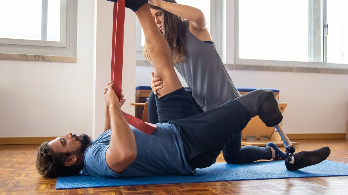
The research supports exercise as rehabilitation for peripheral nerve health, not as nerve repair. Clinical evidence shows positive trends favoring physical therapy after peripheral nerve damage. Those trends were measured by range of motion, muscle power grade and pain [2]. A University of Michigan study looked at nearly 2,900 people who wore wrist activity monitors. Those who spent more time in moderate or vigorous activity were less likely to have peripheral neuropathy [3].
What the research does not show is a path to regrown nerves in people. That difference is the whole point of this article.
How does exercise help peripheral nerves?
Peripheral nerves are long tissue that costs a lot of energy to run. Day after day, they depend on oxygen delivered through the vasa nervorum (the tiny vessels that feed nerves) and on steady metabolic conditions. Both of those are body-wide, not local.
Exercise acts on exactly those systems. Among adults in general, people who are not active enough have a 20% to 30% higher risk of death than people who are active enough [1]. That is a blunt way of saying that the systems exercise touches are the ones that matter most.
What does the research say about exercise and nerve regeneration?
A 2021 review in Heliyon looked at exercise and peripheral nerve regeneration, in animal models and in clinical use. In animal models it reported more axon regeneration and more nerve regrowth into muscle after nerve injury. It also reported better recovery of strength and muscle mass, and higher levels of nerve growth factors [2].
In humans, the same review found clinical evidence of positive trends favoring physical therapy after peripheral nerve damage. That was based on improvement in range of motion, muscle power grade and pain [2]. Those are measures of function. They are not the same as a measured nerve-repair result.
Animal results are not human promises
A result in a rodent model tells you a mechanism is possible. It does not tell you the mechanism works at a useful scale in a person with twenty years of metabolic disease behind them. Here, exercise is used as rehabilitation and as metabolic and blood-vessel conditioning. It is not presented as nerve repair.
Does exercise intensity matter for diabetic neuropathy?
About 30% of people with diabetes have diabetic peripheral neuropathy, and 30% to 40% of those have painful diabetic peripheral neuropathy [3]. Guidelines have recommended physical activity to help prevent diabetic peripheral neuropathy. But they have not said how much or how hard. That is what the Michigan work set out to answer.
They studied nearly 2,900 people wearing wrist activity monitors. More time in moderate or vigorous activity was associated with a lower likelihood of peripheral neuropathy [3]. Association is not causation. And the study measured activity; it did not prescribe it.
Exercise lands better when the terrain is being managed
If the metabolic causes stay active, activity is working against a headwind. That is why we pair movement with metabolic and small-vessel care, instead of offering it as a standalone instruction.
What kinds of exercise help peripheral neuropathy?
The weekly plan at Regenerve combines four parts. Endurance work helps metabolism and blood vessels. Balance and body-awareness training helps stability and function. Then come strength work and daily foot and ankle mobility.
The common mistake is adding intensity too fast. Consistency comes first, then progress, while you watch your symptoms along the way. The full weekly plan and how to adjust it is in exercises for peripheral neuropathy in the feet.
When symptoms flare
Some people feel worse before they feel better. If burning gets worse from one session to the next, the plan gets adjusted instead of pushed through. Usually that means less load and shorter sessions while keeping the same frequency.
Matching the plan to the driver
Neuropathy is a category, not a diagnosis. The best plan depends on what is stressing the nerve. Even when diabetes is present, other causes are often active at the same time. These include medicine effects, alcohol, low nutrients, autoimmune causes, toxic exposures and pinched nerves.
Idiopathic is a label, not an answer
When symptoms are filed as idiopathic, the upstream causes are still worth looking for. Improving the terrain means treating whatever made it fail.
Chemotherapy-induced neuropathy needs pacing
Chemotherapy-induced neuropathy can last and can be sensitive to load. Exercise may still have a role. But the plan has to respect recovery time, and new numbness should be checked before you push through it.
Nutritional and autoimmune drivers change what else you do
When a functional B12 deficiency or a gluten-related problem is driving the injury, exercise alone has limited power to correct it. The shortage has to be treated on its own terms.
Which tests should come before balance training?
Balance training does not help much if the unsteadiness comes from somewhere you have not found. Electrodiagnostic testing, done on site, maps nerve damage. It can tell a body-wide neuropathy apart from a pinched-nerve pattern. Videonystagmography, or VNG, measures inner-ear balance and is a test only.
Frequently asked questions
Can exercise repair damaged nerves?
Animal studies show the idea is plausible, including more axon regeneration and nerve regrowth into muscle. Clinical evidence shows positive trends favoring physical therapy after peripheral nerve damage for range of motion, muscle power grade and pain [2]. Animal findings do not carry over to people automatically, and no one should promise nerve regrowth. See peripheral neuropathy treatments for the feet.
Is walking enough, or does intensity matter?
A University of Michigan study looked at nearly 2,900 people who wore wrist activity monitors. Those who spent more time in moderate or vigorous activity were less likely to have peripheral neuropathy [3]. Intensity appears to matter. But the practical starting point is whatever you can repeat. See peripheral neuropathy of the feet symptoms.
What if exercise makes my feet burn more?
Then the plan needs adjusting, not pushing through. Cut the load, shorten the session, and keep the frequency. That usually keeps the metabolic benefit without setting off the flare. See the neuropathy services offered at Regenerve.
Could my instability be something other than neuropathy?
It can be. Pinched-nerve patterns such as carpal tunnel and sciatica cause overlapping symptoms. Inner-ear balance is a separate question, which VNG testing checks. See carpal tunnel, sciatica and double crush.
What is the best exercise for peripheral neuropathy?
There is no single best exercise. The weekly plan at Regenerve combines endurance work for metabolism and blood vessels, balance and body-awareness training for stability, strength work, and daily foot and ankle mobility. The right mix depends on which cause is doing the most damage. Start with what you can repeat steadily. Then step it up while watching your symptoms.
What should you avoid when exercising with neuropathy?
The most common mistake is adding intensity too fast. Build consistency first, then step it up. If burning gets worse from one session to the next, do not push through it. The plan gets adjusted, usually by cutting load and session length while keeping the same frequency. New numbness is a reason to get checked before you train harder.
Is exercise safe with chemotherapy-induced neuropathy?
Exercise may still have a role. But chemotherapy-induced neuropathy can last and can be sensitive to load. The plan has to respect recovery time and move at a slower pace. If new numbness shows up, get checked before pushing through it. Electrodiagnostic testing, done on site at Regenerve, maps how the nerves are involved.
Can exercise fix neuropathy caused by a B12 deficiency?
Not on its own. When a functional B12 deficiency or a gluten-related problem is driving the nerve injury, exercise has limited power to correct it. The shortage has to be treated on its own terms. Exercise still helps as rehabilitation and as metabolic and blood-vessel conditioning. But it works best alongside care aimed at the actual cause.
Start with the driver
How fast to step up endurance, how much balance work to put first, and what else needs to happen alongside it all depend on which cause is doing the most damage. The free five-question Nerve Damage Score at regenerve.com/assessment is the first step.
World Health Organization, Physical activity (fact sheet), updated 26 June 2024, https://www.who.int/news-room/fact-sheets/detail/physical-activity.
Maugeri G, D’Agata V, Trovato B, Roggio F, Castorina A, Vecchio M, Di Rosa M, Musumeci G, The role of exercise on peripheral nerve regeneration: from animal model to clinical application, Heliyon, 2021;7(11):e08281, doi:10.1016/j.heliyon.2021.e08281, https://pmc.ncbi.nlm.nih.gov/articles/PMC8571504/.
University of Michigan Medical School, New exercise guidelines for neuropathy, https://medresearch.umich.edu/research-news/new-exercise-guidelines-neuropathy.
Numb feet from peripheral neuropathy change driving safety, because the pedal feedback loop depends on feeling you may no longer have. The National Institute of Neurological Disorders and Stroke lists two sensory nerve symptoms that matter here. One is not being able to feel vibration and touch, especially in the hands or feet. The other is loss of position sense.1 Both are exactly what you use to find a pedal and judge how hard you are pressing it.
Why do numb feet affect driving?
Driving is a correction loop. You press, you feel the push back, you adjust. When touch and position sense fade, the correction comes late or lands in the wrong place.
NINDS also notes that loss of position sense can leave a person unable to coordinate complex movements or keep their balance with their eyes shut.1 A footwell is a dark place where your eyes cannot help you.
Symptoms can arrive on any timescale
NINDS states that symptoms of peripheral neuropathy may develop over days, weeks or years. That depends on the type of nerve fibers affected and the type and severity of damage.1 That is why a driving call made two years ago is not a call about today.
Check again after any medicine change, any fall, any new numbness, and any near-miss you explained away. You can read how doctors describe the sensory pattern in peripheral neuropathy of the feet symptoms.
Is it dangerous to drive with neuropathy in your feet?
Does neuropathy slow your braking?
A 2017 case-control study in the Journal of Foot and Ankle Surgery measured brake response time on a computer driving simulator. It compared 25 active drivers with type 2 diabetes and lower-leg neuropathy against 25 active drivers with neither condition.2 The neuropathy group had a slower average brake response time. Abnormally delayed responses also happened more often.
Pressing the wrong pedal
A 2026 study in Transportation Research Interdisciplinary Perspectives used a driving simulation. It compared drivers with and without neuropathy affecting foot feeling and ankle position sense.3 Only the drivers with neuropathy made sudden, powerful, unsafe and unintended accelerations when braking was needed. These were cases of pedal confusion and misapplication.
The authors state that diabetic peripheral neuropathy raises the chance of pressing the wrong pedal. That puts those drivers at higher risk of a crash from unintended acceleration.3 They recommend three countermeasures: drive a manual gearbox instead of an automatic, fit an acceleration limiter, and light the footwell and pedals at night.
When should you stop driving with neuropathy?
These are the patterns to act on, not to shrug off:
Numbness that is spreading or deepening over weeks instead of staying stable.
Having to hunt for the pedal, or being unsure how far down you have pushed it.
Any episode of unintended acceleration, pedal confusion, or a near-miss you cannot explain.
New unsteadiness on uneven ground or in the dark.
A new medication that affects alertness, balance or reaction time.
If driving already feels unreliable, pause it while you get checked. Do not test the question on a public road.
What an evaluation at Regenerve looks at
The useful question is not whether you have neuropathy. It is which cause is behind it and which specific functions have been lost. We do electrodiagnostic testing (EMG/NCS) on site. We sort between metabolic, nutritional, autoimmune, toxic and structural causes.
When balance is part of the picture, we use VNG as a test of inner-ear balance. VNG is a test, not a treatment. Our therapy options include class 4 photobiomodulation, class 3B cold laser and whole-body infrared.
Orthobiologic injections such as PRP and BMAC are offered in selected situations. Qutenza (capsaicin 8% patch) is FDA-approved in adults only for neuropathic pain from postherpetic neuralgia and from diabetic peripheral neuropathy of the feet. It is applied in clinic by a clinician and repeated no more often than every three months. We do not promise nerve regeneration, reversal or cure.
We see patients from the St. Louis region, Missouri and Illinois, at 4477 Woodson Rd #104, St. Louis, MO 63134. Details are on our St. Louis neuropathy treatment page.
Start with your Nerve Damage Score
If your feet have gone quiet, find out what is causing it before you decide anything about driving. The Nerve Damage Score is a free five-question screen.
Can I keep driving if my feet are numb from neuropathy?
That depends on how much feeling and position sense you have lost. It is not something to judge from a good day. NINDS lists loss of vibration and touch feeling and loss of position sense among sensory nerve symptoms. Both affect how well you place and press a pedal. See burning feet and nerve pain.
What is pedal confusion, and is neuropathy really linked to it?
Pedal confusion means pressing the gas when you meant to brake. In a 2026 driving-simulation study, only drivers with diabetic peripheral neuropathy made sudden, powerful, unsafe and unintended accelerations when braking was needed. See the hidden drivers of peripheral neuropathy.
What should I ask about before a driving decision?
Ask about your sensory exam, your position sense, your medicines, and whether a formal driver rehab evaluation fits your case. Bring the specific driving moments that feel unreliable, not a general impression. See Regenerve services and testing.
Does treating the pain make driving safer?
Not by itself. Pain relief and accurate feeling are different things. A treatment that cuts burning does not restore the pressure and position feedback a pedal needs. Qutenza, for example, is approved for nerve pain, not for restoring feeling. See Qutenza (capsaicin 8% patch).
How do you drive a car safely with neuropathy?
Start with an honest check of your feeling and position sense, not a guess from a good day. The 2026 pedal-confusion study recommended three countermeasures: drive a manual gearbox instead of an automatic, fit an acceleration limiter, and light the footwell and pedals at night. Then check again after any medicine change, any fall, any new numbness and any near-miss.
What should you not do if you have neuropathy in your feet?
Do not judge your driving from a good day. Do not explain away a near-miss or a moment of pedal confusion. Do not assume that less pain means safer feet, because pain relief does not restore the pressure and position feedback a pedal needs. And if driving already feels unreliable, do not test the question on a public road. Pause it while you get checked.
Is it harder to drive at night with numb feet?
Yes. Loss of position sense can leave a person unable to coordinate complex movements or keep their balance with their eyes shut. A footwell at night is a place where your eyes cannot help. That is why the 2026 driving study recommended lighting the footwell and pedals at night. It is also why new unsteadiness in the dark is a sign to get checked.
Meyr AJ, Spiess KE. “Diabetic Driving Studies—Part 1: Brake Response Time in Diabetic Drivers With Lower Extremity Neuropathy.” Journal of Foot and Ankle Surgery. 2017;56(3):568–572. https://pubmed.ncbi.nlm.nih.gov/28476387/
Perazzolo M, Esselaar M, Marple-Horvat DE. “Sudden, powerful, unsafe and unintended accelerations: Pedal confusion and misapplication in drivers with diabetic peripheral neuropathy.” Transportation Research Interdisciplinary Perspectives. 2026;38:102129. https://doi.org/10.1016/j.trip.2026.102129
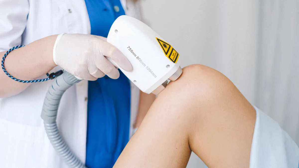
Class 3B cold laser therapy, meaning low-level laser devices in that power range, has been tested against sham treatment in peripheral neuropathy. The results are mixed, not missing. A 50-patient trial in Toronto found a trend toward pain relief that did not reach statistical significance. A 60-patient trial in Iran and a 200-patient trial in India each reported significant improvement in pain and neuropathy scores.1,2,3 No single trial has been large enough, long enough, or similar enough to the others to settle the question.
The second thing worth knowing is who was in those trials. Every one of them enrolled a narrower group of people than the patients who actually walk into a neuropathy clinic. That is the part most articles skip. And it is the part that decides whether a published number applies to you.
This page walks through each trial. It covers the device, the people studied, the exclusion rules as the papers and trial registries state them, and what the design could not measure.
What does “class 3B” mean for a cold laser?
The class is a safety label, not a dose
Laser class is a hazard rating. The federal performance standard at 21 CFR 1040.10 sets the classes. A Class IIIb laser product is one that lets people reach laser radiation, while it runs, above the accessible emission limits of table III-A but not above those of table III-B. A Class IV product goes above table III-B.4 The class describes how dangerous the beam is to an eye or to skin. It says nothing about whether the light helps a nerve.
Two devices in the same class can differ in wavelength and in whether the beam is steady or pulsed. They can differ in how large an area is treated and in how much energy actually reaches tissue. Two devices in different classes can deliver similar energy to similar depth if treatment time and area differ. Class 3B and class 4 are not two schools of therapy. They are two rungs on a safety ladder.
The numbers that matter are often missing from the papers
This is where pooling the research breaks down. A 2016 analysis in Lasers in Medical Science reviewed 74 low-level light therapy and photobiomodulation articles indexed between March 2014 and March 2015. Many left out key settings. Wavelength was missing in 3% of papers, power in 41%, beam area in 40%, irradiance in 43%, pulse frequency in 52%, and radiant energy in 74%.5 Most papers, 73%, gave no information about how light was measured and relied on the maker’s stated values. Only 5% reported a complete set of the settings assessed.
That is not a theory-only complaint. The Toronto trial reports a wavelength of 905 nm and an average power of 0 to 60 mW. But it never states a dose in joules per square centimeter.1 The Iranian trial reports an energy density of 2.5 J/cm². As printed, it also reports a wavelength of 78 nm. That figure is in the extreme ultraviolet range, which no therapeutic laser produces, and the published paper repeats it without correction.2 Neither paper states a laser class at all.
So when someone asks what the trials show for “class 3B,” the honest first answer is this. The trials mostly did not describe themselves in those terms. What they reported was wavelength and, sometimes, power and dose. Reading them means working from those numbers, not from a class label.
The sham-controlled trials that used low-level devices
Toronto, 2004: 50 patients, 905 nm, up to 60 mW
This trial was run at the Diabetic Neuropathy Research Clinic of the Toronto University Health Network between October 2000 and February 2001.1 Fifty patients with painful diabetic sensorimotor polyneuropathy took part: 34 men and 16 women, 88% of them with type 2 diabetes. All received sham therapy for a two-week run-in. They were then randomized to low-intensity laser or continued sham, twice weekly for four weeks. That is eight active sessions in total. The device was a Theralase TLC 5000 at 905 nm with an average power of 0 to 60 mW. It was applied for five minutes to two painful spots on each foot, through a five-prong head covering a 6 cm wide circle of skin.
Who was screened out. Eligible patients were adults aged 18 or older. They had polyneuropathy for at least three months, pain in both feet, and a pain score of at least 4 on the visual analog scale of the Short-Form McGill Pain Questionnaire. Pain medicines and add-on pain medicines were allowed if the regimen was unchanged. The trial excluded patients with unstable medical conditions, naming cancer and active or untreated thyroid disease as examples. It also excluded patients with other nerve diseases that would muddy the neuropathy assessment. Pregnancy, metal implants, alcohol or illicit drug use, and other severe pain that might muddy self-ratings of nerve pain were also exclusions.
What it found. Both groups improved during the sham run-in. The authors put that placebo response at roughly 20% pain reduction. After the four-week treatment, the laser group had an extra drop in weekly mean pain of 1.0 ± 0.4 points, compared with 0.0 ± 0.4 in the sham group. That did not reach statistical significance (P = 0.07). There was no effect on the Toronto Clinical Neuropathy Score, nerve conduction studies, sympathetic skin response, or quantitative sensory testing. The authors concluded that an encouraging trend was seen, but the results did not give enough evidence to recommend the treatment.
What the design could not capture. The authors said this themselves. The study ran over a short period with a fairly small number of patients. Any benefit of low-intensity laser may be limited to small nerve fibers, which are hard to assess accurately. And clearer results might have come from a larger sample and more frequent or longer treatment. They also recorded that no significant side effects occurred. There was no concern about interactions in patients on several medicines. So the treatment could be offered safely to patients with a wide range of other diseases.
Kermanshah, 2013: 60 patients, twice weekly for a month
Sixty patients with diabetic sensorimotor polyneuropathy took part at a diabetes research center in Kermanshah, Iran.2 They were matched on sex, age, body mass index, type and length of diabetes, and length of pain. Then a coin toss randomized them to laser or sham. Participants were 77.7% female, with a mean age of 56.1 years. Diabetes duration ranged from 24 to 360 months, and neuropathy duration from 4 to 96 months. Treatment was twice weekly for five minutes over one month, about eight sessions.
Who was screened out. Eligible patients were older than 18 and diagnosed with polyneuropathy for more than three months. They had pain in both feet and a visual analog score of at least 4. Patients on medicine could join if the regimen had not changed in the four weeks before the study. The trial excluded patients with cancer, patients with thyroid disease who were not taking medicine for it, and patients with nerve disorders that could get in the way of evaluation. Pregnant women, patients with metal objects in the body, and patients using alcohol or psychotropic drugs were also removed.
What it found. In the laser group, visual analog and Toronto Clinical Neuropathy scores improved significantly from baseline at both two and four weeks (P < 0.0001). The difference against sham was also significant (P < 0.0001). The sham group showed no significant change on either measure. Within the laser group, neuropathy scores at four weeks were not significantly better than at two weeks. So most of the change appeared to come early and then level off.
What the design could not capture. Follow-up ended four weeks after a one-month course. So nothing in this trial speaks to how long the effect lasts. The report gives an energy density but no power output, no treatment area, and no laser class. That makes the protocol hard to repeat or to pool with other studies.
Manipal, 2025: 200 patients, helium-neon at 632.8 nm
This is the largest of the low-level laser trials in this condition. Two hundred people with type 2 diabetes and peripheral neuropathy were randomized, 100 per arm, at a center in Manipal, India.3 The treatment arm received photobiomodulation for ten days. It used a low-level helium-neon laser at 632.8 nm and 3.1 J/cm² for nine minutes across the top and sole of the foot. The control arm received sham laser. Patients were assessed on day 0 and four weeks after treatment. The registered protocol also lists a second low-level device. It was a Thor laser at 660 and 850 nm with a power density of 50 to 150 mW/cm², applied behind the knee (popliteal fossa) and over the neck of the fibula. The published report describes the helium-neon part.6
Who was screened out. The trial registration lists inclusion as people aged 30 to 70 with type 2 diabetes and peripheral nerve pain who were willing to consent.6 It excluded patients with any other nerve disorder or other illnesses (morbid conditions). It excluded patients with another metabolic neuropathy, giving thyroid disorders as the example. It excluded pregnant women and patients having chemotherapy or radiation. And it excluded patients with diabetic peripheral neuropathy who had a foot ulcer.
What it found. Four weeks after treatment, the treatment group showed significant change in several measures, all at P < 0.001. These were serum neuron specific enolase and calcitonin gene-related peptide, the Michigan Neuropathy Screening Instrument score, vibration perception threshold, numeric pain rating, and the Norfolk quality-of-life score. Nerve growth factor did not change (P = 0.937). The authors concluded that serum neuron specific enolase and calcitonin gene-related peptide may be good markers of how well photobiomodulation reduces nerve pain and other symptoms.
What the design could not capture. Only the participants were blinded, not the assessors. That is weaker than the double-masked design used in Toronto. The exclusion list removes the two groups a neuropathy clinic sees all the time. These are people whose nerve injury has more than one cause, and people whose feet have already broken down. And ten days of treatment with a four-week check cannot say anything about a year from now.
What happens when these studies are pooled
A 2019 systematic review searched PubMed, Web of Science, CINAHL and Cochrane. It screened 627 records and included six studies, randomized and non-randomized. Its outcomes were pain score, nerve conduction velocity and quality-of-life measures.7 It concluded that the evidence obtained shows low level laser therapy has a positive effect in controlling diabetic nerve pain. Six studies of mixed design is a small and uneven base, and the review says so by reporting its own numbers.
A 2025 review in Lasers in Medical Science looked at 23 clinical and preclinical studies published between 2015 and 2025. It focused on dose.8 It reported the most consistent treatment effects at wavelengths of 630 to 670 nm and 808 to 904 nm. Those effects also came with fluences of 3 to 10 J/cm², output power between 45 and 100 mW, and protocols of at least 12 sessions. It described photobiomodulation as a promising, safe and effective add-on therapy for diabetic neuropathy. And it concluded that the lack of standard dosing across studies remains a major challenge.
Two details in that set of settings are worth pausing on. An output power of 45 to 100 mW sits inside the class 3B range, not the class 4 range. And the session count matters. The Toronto and Kermanshah trials each gave about eight sessions, and the Manipal trial ten. That is fewer than the twelve-session floor later tied to more consistent effect.
The class 4 trials, and why they are a separate question
Older adults with type 2 diabetes
The trial most often quoted in cold laser marketing used a class IV device, not a class 3B one. It was a pilot randomized, double-masked, sham-controlled study of 40 adults aged 60 and above. They had type 2 diabetes and painful diabetic peripheral neuropathy confirmed by nerve conduction velocity. They were recruited from the Department of Geriatric Medicine at AIIMS in New Delhi between April 2016 and September 2017.9 Participants were randomized 1:1 to deep tissue laser therapy or sham. Sessions were twice weekly for four weeks and then weekly for eight weeks, sixteen sessions in total. Both arms continued standard care. So the trial tested laser as an add-on to usual care, not as a replacement for it.
The device was a Class IV medical laser. It delivered a blend of 980 nm and 810 nm light in an 80:20 optical power ratio, at 2 W continuous wave, with a starting combined irradiance of 0.8 W/cm². It covered a sole area of about 120 to 200 cm². Class 3B units run at much lower output. So this trial does not tell you what a class 3B device would do.
Who was screened out. The paper excluded patients with type 1 diabetes and open wounds on the legs or feet. It excluded psychotic, mood or nerve disorders that could get in the way of the assessments. It also excluded patients with a life expectancy of less than three years and any cancer treated with chemotherapy or radiotherapy in the previous two years. Patients were recruited no matter how long they had had neuropathy, so both new and long-standing cases were included.
What it found. Pain fell in both arms, and significantly more in the active arm. On the Pain Disability Questionnaire, the active group fell 60% (P < 0.001), against 28% in sham (P < 0.01). Quality of life improved significantly in the active group (P < 0.001) but not in sham (P = 0.194). Timed Up and Go times improved in both groups, and the improvement was 16% greater in the active group. But the authors reported that the ANOVA interaction for that difference was not statistically significant (P > 0.05). That correction matters. The function result was not a positive finding, and it should not be quoted as one.
What the design could not capture. The authors listed their own limits. There was no data on how long patients had had neuropathy, and disease duration varied among participants. Some comparisons lacked statistical power. And as a pilot, it needs to be confirmed in a multicenter trial with a larger group and longer follow-up.
Chemotherapy-induced neuropathy
Two class IV trials tested a different cause. One was a randomized, double-blind, sham-controlled cross-over trial at the University of Minnesota.10 In it, 70 patients with chemotherapy-induced peripheral neuropathy got 30-minute sessions three times weekly for six weeks. They received either a class IV laser at 800 to 970 nm, 6.75 to 12 W per treatment zone, or a sham ceramic heat probe. Eligibility here was unusually broad. Any adult with self-reported peripheral neuropathy and past chemotherapy could enroll. That included patients diagnosed with neuropathy before chemotherapy and patients already on drug treatment for it, as long as they did not change therapy during the study. The single stated exclusion was active cytotoxic cancer treatment within 30 days of enrollment.
Treated patients had a mean drop in modified total neuropathy score of 6.8 points, 52.6%, at eight weeks (P < 0.001). Sham patients changed by +0.2 points, 1.5% (P = 0.44). Adding physiotherapy to the laser did not improve the result over laser alone (difference 0.1 points, P = 0.85). But the group was entirely female and 98% non-Hispanic white, because it was drawn from a gynecologic oncology clinic. Broad on other diseases, narrow on who the patients were.
The NEUROLASER pilot in Belgium took the opposite approach. It tested prevention, not treatment.11 Thirty-two breast cancer patients getting taxane chemotherapy were analyzed, after 54 were randomized. It used a class IV device at 905 and 808 nm, 0.168 W/cm² and 4 J/cm², twice weekly. The exclusions were long. They included prior neuropathy from another condition and being on a stable dose of neuropathy medicine such as duloxetine or pregabalin. They also included cancer that had spread, pausing chemotherapy for more than two cycles, missing more than two laser sessions in a row, and any cut in chemotherapy dose. Neuropathy scores rose significantly over time in both arms (P < 0.001), and the difference between arms was not significant. Still, quality of life, the six-minute walk at follow-up (P = 0.035) and pain at follow-up (P = 0.058) favored the treated group. The authors named the small sample as their main limit.
Who did the cold laser studies leave out?
Line the exclusion lists up and a pattern appears. Across these six trials, the following people were kept out of at least one, and usually several. Patients with type 1 diabetes. Patients with an open foot wound or ulcer. Patients with any other nerve disease. Patients with a second metabolic cause such as thyroid disease. Patients with cancer, or on chemotherapy or radiation, or with a life expectancy under three years. Patients with other severe pain that would muddy a pain questionnaire. Patients whose medicine regimen had changed recently. And, in the largest trial, patients over 70 or under 30, and anyone with unnamed “other morbid conditions.”
These are reasonable scientific choices. Removing competing explanations is how a trial isolates one variable. But the result is that the published numbers describe a cleaner patient than the one in the exam room. Picture a person with type 2 diabetes, insulin resistance, a thyroid disorder, a healed ulcer, a recent change in gabapentin and eight years of symptoms. That person would have been ruled out of most of this research.
The conclusion cuts both ways, and it should. A trend that missed statistical significance in 50 carefully chosen patients does not prove that low-level laser fails in patients who were screened out. And a significant result in 200 carefully chosen patients does not prove that it will help someone the trial would have excluded. What the exclusions show is that the question has not been asked in the complex patient at all.
What can the cold laser trials not tell you?
Four gaps run across the whole set.
Duration. Active treatment ran four weeks in Toronto, one month in Kermanshah, ten days in Manipal, twelve weeks in New Delhi and six weeks in Minnesota. The longest follow-up in any of them was 16 weeks. Nothing here measures how long the effect lasts at a year.
Combination and sequence. Only the New Delhi trial clearly gave laser on top of ongoing standard care. Only the Minnesota trial tested adding a second treatment, physiotherapy, and that did not improve the result over laser alone. None of them tested light inside a step-by-step program that first finds and corrects the metabolic, nutritional, autoimmune or toxic cause. A single-treatment trial cannot answer a combination question.
Outcome measures. Standard nerve conduction studies mainly test large, coated (myelinated) fibers. The Toronto authors expected no change in those tests and got none. They raised the chance that any benefit lies in small fibers that these tests do not measure well. So a trial can report a null objective result while missing the fibers that carry burning and temperature feeling.
Dose. Settings were reported unevenly, and in two of these papers incompletely or implausibly. So the trials cannot be added together into one answer about one treatment. They are several different treatments sharing a label.
Does Medicare cover laser treatment for neuropathy?
Medicare maintains national coverage determination 270.6, Infrared Therapy Devices, effective for services on and after October 24, 2006.12 It states that the use of infrared or near-infrared light or heat, including monochromatic infrared energy, is non-covered for the treatment of diabetic or non-diabetic peripheral sensory neuropathy and of wounds or ulcers of the skin or subcutaneous tissues, including pain arising from those conditions. The determination it followed was proposed in the CAG-00291N decision memorandum dated July 26, 2006.13
Two things are worth reading carefully. First, the NCD’s own description of the device category is an array of juxtaposed infrared diodes affixed to a flexible pad held in skin contact — a diode pad, not a laser. Second, a coverage determination is a payment decision made under the reasonable-and-necessary standard of the Social Security Act. It is not a clinical finding, and it does not report a trial result. Patients sign an Advance Beneficiary Notice for services a payer does not cover. Whether something is covered and whether something helps are two different questions, and a page that treats them as one is misleading its reader.
Why the driver matters more than the device
A laser cannot correct a nutrient shortage, an autoimmune process or a toxic exposure. Before adding a light-based therapy to a plan, the more useful question is which cause is damaging the nerve right now.
That is why our workup starts with finding the cause, whether metabolic, nutritional, autoimmune, toxic or structural, instead of treating “neuropathy” as one condition. You can read how we sort those apart in the hidden drivers of peripheral neuropathy.
A 2025 review of laser therapy for nerve pain searched four databases for English-language studies published between January 2000 and June 2025. It covered both low-level and high-intensity laser therapy, across conditions including postherpetic neuralgia and diabetic neuropathy.14 The breadth of that research is exactly why a single device label such as “3B” does not predict a result.
How we use class 3B cold laser at Regenerve
Regenerve offers class 3B cold laser alongside class 4 photobiomodulation and whole-body infrared. We use them as parts of a physician-directed plan. That comes after on-site electrodiagnostic testing and a metabolic workup have found what is driving the nerve injury. And it goes alongside the metabolic and nutrition work that treats that cause. We do not use light as a stand-alone treatment for nerve damage.
We do not promise nerve regeneration, reversal or cure. We do not publish a success rate for our protocol. Our full list of testing and therapy options is on the Regenerve services page.
What we tell patients before starting
The talk covers four things. First, what the published trials for your cause actually showed, including where they were negative. Second, whether you would have qualified for those trials, and if not, what that does and does not mean. Third, the safety record, which across these studies was benign. The Toronto trial reported no significant side effects and no concern about interactions in patients on several medicines. The Minnesota trial reported no complications among treated patients. And fourth, a set point at which we stop and reassess instead of going on forever.
“My ethos is to treat all of my patients as I would my own family, with the goal of giving them back their quality of life.” — Dr. Gurpreet Singh Padda, MD, MBA, MHP
We see patients from the St. Louis region, Missouri and Illinois, at 4477 Woodson Rd #104, St. Louis, MO 63134, minutes from St. Louis Lambert International Airport. Call (314) 886-5739 or text (314) 886-5902.
Start with your Nerve Damage Score
If you are weighing cold laser or any other therapy, start by finding out which cause is doing the most damage. The Nerve Damage Score is a free five-question screen that helps focus the first visit.
Does class 3B cold laser therapy work for neuropathy?
The sham-controlled evidence for low-level devices in this power range is mixed. A 50-patient trial in Toronto found a trend that missed statistical significance at P = 0.07. A 60-patient trial in Iran and a 200-patient trial in India each reported significant improvement in pain and neuropathy scores. All three enrolled selected groups. So the results describe those groups, not every patient. See peripheral neuropathy treatments for the feet.
Is cold laser the same thing as infrared therapy for neuropathy?
No. Medicare’s national coverage determination 270.6 describes infrared therapy devices as an array of infrared diodes on a flexible pad held against the skin. That device category is not covered for diabetic and non-diabetic peripheral sensory neuropathy. A laser differs in wavelength, coherence, power and delivery. And a coverage decision is a payment judgment, not a trial result. See alternative medicine approaches to peripheral neuropathy.
Should I have nerve testing before trying laser therapy?
Testing first tells you which fibers are involved. It shows whether the pattern starts in the longest nerves, sits in one spot, or involves small fibers. That changes what is worth trying. Nerve conduction studies mainly test large, coated fibers. So they can read normal when small fibers are the problem. That is also why a trial using them as an endpoint can miss an effect. See small fiber neuropathy symptoms and testing.
Does laser therapy help if my neuropathy is not diabetic?
Most of the low-level laser evidence was gathered in diabetic polyneuropathy. Two class 4 trials studied chemotherapy-induced neuropathy. One reported a large drop in neuropathy scores. The other found no significant difference from sham on its main endpoint. Autoimmune, nutritional and toxic causes have not been tested this way, and the plan should still target the cause doing the damage. See diabetic peripheral neuropathy treatment in St. Louis.
What are the drawbacks of laser therapy for neuropathy?
The main drawbacks are gaps in the evidence, not harm. The longest follow-up in these trials was 16 weeks, so nobody knows how long a benefit lasts. Doses were reported so unevenly that the trials cannot be added together. And most of them screened out complex patients. Light also cannot correct the nutrition, autoimmune or toxic cause that is damaging the nerve.
How many laser sessions does neuropathy treatment take?
The published trials used fewer sessions than you might expect. The Toronto and Kermanshah trials each gave about eight. The Manipal trial treated for ten days, and the New Delhi class 4 trial used sixteen. A 2025 dosing review tied more consistent effects to protocols of at least twelve sessions. At Regenerve we set a point to stop and reassess instead of going on forever.
Is cold laser therapy safe for neuropathy?
Across these studies the safety record was benign. The Toronto trial reported no significant side effects and no concern about interactions in patients taking several medicines. The Minnesota trial reported no complications among treated patients. Laser class is a hazard label for the eyes and skin, not a dose. We use cold laser as one part of a physician-directed plan, never on its own.
Sources
Zinman LH, Ngo M, Ng ET, Nwe KT, Gogov S, Bril V. “Low-intensity laser therapy for painful symptoms of diabetic sensorimotor polyneuropathy: a controlled trial.” Diabetes Care. 2004;27(4):921–924. https://doi.org/10.2337/diacare.27.4.921
Bashiri H. “Evaluation of low level laser therapy in reducing diabetic polyneuropathy related pain and sensorimotor disorders.” Acta Medica Iranica. 2013;51(8):543–547. https://acta.tums.ac.ir/index.php/acta/article/view/4395
Anju M, Ummer Velladath S, Arun Maiya G, Hande M. “A single blinded randomized controlled trial assessing the effect of photobiomodulation therapy on neuron specific biomarkers in type II diabetes mellitus patients with peripheral neuropathy.” Diabetes Research and Clinical Practice. 2025;222:112087. https://doi.org/10.1016/j.diabres.2025.112087
Hadis MA, Zainal SA, Holder MJ, Carroll JD, Cooper PR, Milward MR, Palin WM. “The dark art of light measurement: accurate radiometry for low-level light therapy.” Lasers in Medical Science. 2016;31(4):789–809. https://pmc.ncbi.nlm.nih.gov/articles/PMC4851696/
Clinical Trials Registry – India. “Effect of Low Level Laser Therapy on Neuropathic Pain Specific Markers in Diabetic Peripheral Neuropathy — A Single Blinded Randomised Control Trial.” CTRI/2018/04/013540, registered April 27, 2018. WHO International Clinical Trials Registry Platform record. https://trialsearch.who.int/Trial2.aspx?TrialID=CTRI/2018/04/013540
Anju M, Saleena Ummer V, Arun G Maiya, Manjunath Hande. “Low level laser therapy for the patients with painful diabetic peripheral neuropathy — A systematic review.” Diabetes & Metabolic Syndrome. 2019;13(4):2667–2670. https://doi.org/10.1016/j.dsx.2019.07.035
de Souza V, da Palma Cruz M, Chaves Bittencourt C, et al. “Dosimetric parameters and clinical outcomes of photobiomodulation in diabetic neuropathy: a concise review.” Lasers in Medical Science. 2025;40:436. https://doi.org/10.1007/s10103-025-04701-7
Chatterjee P, Srivastava AK, Kumar DA, Chakrawarty A, Khan MA, Ambashtha AK, Kumar V, De Taboada L, Dey AB. “Effect of deep tissue laser therapy treatment on peripheral neuropathic pain in older adults with type 2 diabetes: a pilot randomized clinical trial.” BMC Geriatrics. 2019;19:218. https://pmc.ncbi.nlm.nih.gov/articles/PMC6689877/
Argenta PA, Ballman KV, Geller MA, Carson LF, Ghebre R, Mullany SA, Teoh DGK, Winterhoff BJN, Rivard CL, Erickson BK. “The effect of photobiomodulation on chemotherapy-induced peripheral neuropathy: A randomized, sham-controlled clinical trial.” Gynecologic Oncology. 2017;144(1):159–166. https://doi.org/10.1016/j.ygyno.2016.11.013
Lodewijckx J, Robijns J, Claes M, et al. “The use of photobiomodulation therapy for the prevention of chemotherapy-induced peripheral neuropathy: a randomized, placebo-controlled pilot trial (NEUROLASER trial).” Supportive Care in Cancer. 2022;30(6):5509–5517. https://pmc.ncbi.nlm.nih.gov/articles/PMC8935622/
Yes, peripheral neuropathy can improve. How much depends on which cause is damaging the nerve and how long that cause has been at work. Cleveland Clinic states that some types of neuropathy can be stopped or improved when they are found and treated quickly.1 For damage that has built up over years, real goals shift toward less burning, better protective feeling and safer walking.
Peripheral neuropathy is the term for any nerve damage outside the brain and spinal cord.1 That one definition is why the honest answer varies so much from person to person. The label tells you where the injury is. It does not tell you what is causing it.
Key takeaways
Neuropathy is a category, not a diagnosis. What can improve depends on the driver, not the label.
Most stalled cases are cases where the cause was never found. Treatment aimed at the wrong problem tends to stall.
Testing narrows the question. We do electrodiagnostic testing (EMG and nerve conduction studies) on site. It helps show which nerve fibers are involved.
Timing matters. A cause caught early is a different problem from one that has been at work for a decade.
For long-standing damage, the goals change. The targets are less burning, more reliable feeling and steadier walking, not nerve regrowth.
Why does neuropathy improve for some people and not others?
Two people can describe the same burning feet and the same numbness and have totally different problems underneath. One may have an active metabolic problem. Another may have a nutrient shortage, an autoimmune process, a toxic exposure or a pinched nerve.
That is why the useful question is not “how bad is my neuropathy.” It is “what is still injuring my nerves right now, and can that be reduced.”
The five driver categories we work through
Metabolic drivers, including the nerve injury seen in diabetes
Nutritional drivers, including B12 deficiency
Autoimmune and infectious drivers
Toxic and environmental drivers
Structural and compressive contributors
Medicine that turns down the signal does not repair the wiring. If the cause is still active, symptoms tend to come back whenever the dose is lowered.
What does improvement in neuropathy look like?
Improvement is not one number. It is a set of changes that matter for daily life and for safety.
Goals that are realistic to discuss
Less burning at night and fewer hot-coal sensations
Better protective feeling, so you notice pressure before the skin breaks down
Less numbness and more reliable feedback from the feet, which helps you walk more steadily
Fewer electric, shooting misfires
Holding steady, once exposure to the cause has been cut
What we do not promise
We do not promise nerve regeneration, reversal or a cure. When nerve injury has had years to build up, the honest goal is to heal the nerve’s environment and improve function. It is not a return to how your feet felt before symptoms started.
How is peripheral neuropathy diagnosed?
In diabetic peripheral neuropathy, StatPearls notes that peripheral neuropathy is mainly diagnosed clinically.2 That means your history, your symptom pattern and the exam carry much of the weight. Testing answers specific questions the exam raises.
Mapping before choosing a treatment
On-site electrodiagnostic testing (EMG and nerve conduction studies) to check nerve function
Cause workup across metabolic, nutritional, autoimmune, toxic and structural causes
A plan that follows the finding, not a fixed sequence applied to everyone
Can diabetic neuropathy improve?
Diabetic peripheral neuropathy is a metabolic and small-vessel problem that happens to show up in the feet. How much can improve depends mostly on how much of that metabolic injury is still active. That is why sugar control and blood-vessel support sit inside the treatment plan, not beside it. Our approach for patients in this situation is on the diabetic peripheral neuropathy treatment page.
Why the timeline matters
High blood sugar drives a chemical reaction in the body. It is related to how sugar browns under heat. The longer that process runs unchecked, the more of the nerve’s support system it harms. You can read how that process damages nerve tissue in our article on glycation and nerve damage.
Where light-based and energy-based therapies fit
Class 4 photobiomodulation, class 3B cold laser and whole-body infrared are used at Regenerve as part of a broader plan aimed at the nerve environment. They are not a stand-alone answer, and they are not a substitute for identifying the driver.
When light therapy is added without knowing what is injuring the nerve, the result is a gamble. When it is added to a plan that also cuts the active cause, it has a clear job inside that plan.
When the diagnosis comes back “idiopathic”
Many people arrive with an idiopathic label. That usually means the cause was not found during the last workup. That is a fork in the road, because a cause that has not been found cannot be cut back.
Small fiber damage, nutrient shortage, gluten-related problems, autoimmune processes and toxic exposures are all worth working through before you accept that there is no cause. Some of these turn up on a second look because the first workup was aimed elsewhere.
Regenerve in the St. Louis region
Regenerve treats peripheral neuropathy for patients across the St. Louis region, Missouri and Illinois, including Edwardsville, Glen Carbon, Collinsville, Troy and Maryville. The clinic is at 4477 Woodson Rd #104, St. Louis, MO 63134, minutes from St. Louis Lambert International Airport.
Call (314) 886-5739 or text (314) 886-5902, or email info@regenerve.com. Hours are Monday through Thursday 9 a.m. to 4 p.m. and Friday 9 a.m. to 1:30 p.m.
Your next step
If you want a clear starting point, take the free five-question Nerve Damage Score. It shows which group of causes most likely applies to you. Your answers guide what we check first.
Can peripheral neuropathy improve if both of my feet are affected?
It can. An even, feet-first pattern is common when a metabolic or nutrition cause is at work. Finding that cause is what changes the outlook. The workup, including on-site electrodiagnostic testing, is on our neuropathy services page.
My neuropathy was called idiopathic. Does that mean nothing can be done?
Why do my symptoms feel worse than my test results suggest?
Small nerve fibers carry burning, pins and needles and temperature feeling. They can be injured while routine nerve conduction testing still looks close to normal. See small fiber neuropathy symptoms and testing.
What can I realistically expect for burning feet at night?
Burning at night is one of the symptoms most likely to respond when the cause is treated. The timeline varies, and no one can promise a specific result. See burning feet and nerve pain.
Can peripheral neuropathy ever go away?
For some types, yes. Cleveland Clinic states that some types of neuropathy can be stopped or improved when they are found and treated quickly.1 What decides it is which cause is injuring the nerve and how long it has been at work. When damage has built up over years, the real goals shift to less burning, better protective feeling and safer walking. They are not a return to how your feet felt before.
Can neuropathy be cured?
We do not promise a cure, nerve regeneration or reversal. Neuropathy is a category, not a diagnosis. So what can change depends on the cause underneath it. When a cause is caught early and cut back, holding steady and real symptom relief are fair to discuss. When nerve injury has had years to build, the honest goal is a healthier nerve environment and better daily function.
What makes peripheral neuropathy worse?
A cause that is still active and still injuring the nerve. That can be a metabolic problem such as diabetes, or a nutrient shortage such as low B12. It can be an autoimmune, infectious or toxic process, or a pinched nerve. In diabetes, high blood sugar drives a chemical reaction related to how sugar browns under heat. The longer it runs unchecked, the more of the nerve’s support system it harms.
What are the ways to treat peripheral neuropathy?
Treatment starts with finding what is injuring the nerve. We work through metabolic, nutritional, autoimmune, toxic and structural causes. We run EMG and nerve conduction studies on site. Then we build the plan around what we find. In diabetes, sugar control and blood-vessel support sit inside the plan. Class 4 photobiomodulation, class 3B cold laser and whole-body infrared each have a clear job within that plan. None is a stand-alone answer.
Sources
Cleveland Clinic, “Peripheral Neuropathy,” Cleveland Clinic health library, last updated July 16, 2026, https://my.clevelandclinic.org/health/diseases/14737-peripheral-neuropathy
Bodman MA, Dreyer MA, Varacallo MA, “Diabetic Peripheral Neuropathy,” StatPearls, Treasure Island (FL): StatPearls Publishing, last updated February 25, 2024, https://www.ncbi.nlm.nih.gov/books/NBK442009/
In diabetic peripheral neuropathy, alpha-lipoic acid probably has little or no effect on neuropathy symptoms compared with placebo. That is what a 2024 Cochrane review found. It pooled three placebo-controlled randomized trials in 816 adults with type 1 or type 2 diabetes. The mean difference was −0.16 points on symptom scores at six months. There was also low-certainty evidence of little or no effect on impairment [1]. An American Family Physician summary of the same review showed the same picture at both six and 24 months [2].
That does not make alpha-lipoic acid harmful. It also does not answer every neuropathy question. It means this: if you want a clear change in burning or numbness from diabetic peripheral neuropathy, the trials do not support expecting it from this supplement alone.
Does alpha-lipoic acid work for neuropathy?
Trial wording can sound slippery, so here it is in plain terms. The people studied were adults with diabetes and diabetic peripheral neuropathy. Their mean age was 57.8 years. They were treated for at least six months at doses between 600 and 1,800 mg per day. On average, people on alpha-lipoic acid did about the same as people on placebo [1].
The reviewers also found little or no difference in side effects that made people stop treatment [1][2]. So the honest summary is not that this supplement is dangerous. It is that it is unlikely to change how your feet feel.
What the review actually measured
The main outcome was the change in neuropathy symptoms six months after randomization. It was measured with proven tools such as the Total Symptom Score. Other outcomes were symptoms at 24 months, impairment at six and 24 months, and side effects that made people stop [1].
The reviewers lowered their certainty because people dropped out of follow-up and the results were loose, mostly for impairment [1]. That difference matters. The finding is not that the supplement was proven useless. It is that no benefit showed up, on evidence of moderate and low certainty.
Why doesn’t one supplement fix neuropathy?
Peripheral neuropathy is a category, not a diagnosis. Nerve injury can come from metabolic stress, low nutrients, autoimmune attack, toxic exposure or a pinched nerve. Those causes do not respond to the same fix.
That is why the first question in our clinic is not which supplement to add. It is which cause is doing the most damage right now. You can read how we handle the causes that often get missed in the hidden drivers of peripheral neuropathy.
When the driver is metabolic
When the main cause is metabolic, the plan has two goals at once. First, lower the ongoing metabolic strain on nerve fibers and their tiny blood vessels. Second, support the nerve’s environment while that strain comes down. A supplement added on top of an unchanged metabolic picture is swimming against the current.
Low nutrients, gluten-linked patterns, toxic and environmental exposures, autoimmune attack and chemotherapy nerve injury all cause nerve symptoms. None of them were the group studied in the alpha-lipoic acid trials.
A label of idiopathic usually means the cause has not been found yet. It does not mean there is no cause. That is a reason to keep looking, not a reason to cycle through supplements.
What should you do instead of guessing with supplements?
We do electrodiagnostic testing (EMG and nerve conduction studies) on site. So we read your nerve conduction results alongside your history, not as a separate report weeks later. We pair that with a metabolic and nutrition workup to check whether the terrain has what nerves need.
When in-clinic therapy fits, our confirmed options include class 4 photobiomodulation, class 3B cold laser and whole-body infrared. We use these inside a plan matched to your cause, not as a standalone answer. We do not promise nerve regeneration or reversal of established nerve damage.
Should you keep taking alpha-lipoic acid?
Some patients already take it and tolerate it well. Our position is that it can stay as one part of metabolic and nutrition support. That holds as long as it does not push aside the work that actually changes the cause. The trial evidence should set your expectations for what it will do.
Frequently asked questions
Does alpha-lipoic acid help diabetic peripheral neuropathy?
Probably not enough for you to notice. A 2024 Cochrane review of three placebo-controlled trials in 816 adults with diabetes looked at this. It found that alpha-lipoic acid probably has little or no effect on neuropathy symptoms at six months, and may have little or no effect on impairment [1]. Our approach when diabetes is the cause is on our diabetic peripheral neuropathy treatment page.
Does taking it for longer work better?
Not according to the published record. An American Family Physician summary of the same Cochrane review found little or no effect compared with placebo. That held for symptoms and impairment at both six and 24 months [2]. When low nutrients are the real problem, fixing that shortage is a different question. See functional B12 deficiency and neuropathy.
Do these results apply to me if my neuropathy is not from diabetes?
No. The trials enrolled adults with type 1 or type 2 diabetes and diabetic peripheral neuropathy [1]. So the finding cannot be carried over to autoimmune, toxic, nutritional or idiopathic causes. Burning pain that lasts when routine nerve conduction testing looks close to normal points somewhere else. See small fiber neuropathy symptoms and testing.
If not a supplement, what is the first useful step?
Finding which cause is active, then matching the testing to that pattern. We do electrodiagnostic testing (EMG and nerve conduction studies) on site. We read it alongside a metabolic and nutrition workup, not one change at a time. See our neuropathy services.
How much alpha-lipoic acid was tested for neuropathy?
The trials in the 2024 Cochrane review gave adults with diabetic neuropathy between 600 and 1,800 mg a day for at least six months [1]. Even across that range, the average result was close to placebo. So the dose matters less than your expectations. At the amounts studied, this supplement is unlikely to change how your feet feel on its own.
Is there a downside to taking alpha-lipoic acid?
The reviewers found little or no difference versus placebo in side effects that made people stop treatment [1][2]. The bigger downside is time. If alpha-lipoic acid takes the place of finding the cause of the nerve damage, that cause keeps working. If you already take it and tolerate it well, it can stay as one part of a plan, not the plan.
What is the number one supplement for neuropathy?
There isn’t one. Peripheral neuropathy is a category, not a diagnosis. Metabolic stress, low nutrients, autoimmune attack, toxic exposure, and nerve compression all injure nerves. They do not respond to the same fix. That is why our first question is which cause is doing the most damage right now, not which supplement to add.
Can alpha-lipoic acid regenerate nerves?
The trial record does not show it. The Cochrane review found little or no effect on symptoms at six months. It also found low-certainty evidence of little or no effect on nerve impairment [1]. We do not promise nerve regeneration or reversal of established damage from any treatment. The goal is to cut what is still injuring the nerve and support the conditions in which function can improve.
Start with the Nerve Damage Score
The Nerve Damage Score is a free five-question check. It helps find which cause is most likely behind your symptoms. That way your workup starts with a direction instead of a guess.
Regenerve, 4477 Woodson Rd #104, St. Louis, MO 63134. Serving the St. Louis region, Missouri and Illinois, minutes from St. Louis Lambert International Airport.
Sources
Baicus C, Purcarea A, von Elm E, Delcea C, Furtunescu FL. “Alpha-lipoic acid for diabetic peripheral neuropathy.” Cochrane Database of Systematic Reviews, 2024, Issue 1, Art. No.: CD012967. Published 11 January 2024. DOI: 10.1002/14651858.CD012967.pub2. https://www.cochranelibrary.com/cdsr/doi/10.1002/14651858.CD012967.pub2/full — cited for the three included placebo-controlled trials, 816 adult participants with type 1 or type 2 diabetes, mean age 57.8 years, doses of 600–1,800 mg per day, the −0.16-point symptom difference and −1.02-point impairment difference at six months, the outcome definitions, and the certainty downgrades for loss to follow-up and imprecision.
Abuali SM, Frasca DJ. “Alpha-Lipoic Acid for Diabetic Peripheral Neuropathy.” American Family Physician, February 2025;111(2):119–120. https://www.aafp.org/afp/2025/0200/mbtn-alpha-lipoic-acid-diabetic-peripheral-neuropathy — cited for little or no effect versus placebo on symptom reduction and impairment at six and 24 months, and little or no difference in adverse events.
Autonomic neuropathy is damage to the nerves that run the body’s automatic functions. It goes unnoticed for years because its symptoms show up looking like organ problems. Feeling lightheaded on standing, feeling full after a few bites, a change in sweating, bladder trouble, and pupils slow to adjust between light and dark are all known autonomic symptoms [1]. Each one usually gets sent to a different specialist before anyone asks whether one set of nerves ties them together.
These nerves control heart rate and blood pressure, digestion, bladder and sexual function, sweating and body heat, and the pupils [1]. When those signals fail, the body looks like it has several unrelated problems at once.
Why is autonomic neuropathy so often missed?
Autonomic symptoms are scattered by nature. The autonomic nervous system reaches nearly every organ. So damage to it causes complaints in nearly every clinic, and each complaint makes sense on its own.
That is why diagnosis takes so long. The symptoms are not vague. They are spread out. And spread-out symptoms get split up instead of added together.
Can autonomic neuropathy make you dizzy when you stand up?
Nerve damage can make you feel dizzy or faint when you stand up or exert yourself. It can also cause a fast heart rate or sudden heart-rate changes [1]. In some people it blocks the chest pain that would normally warn of a heart problem [1].
Why it is usually blamed on dehydration
Feeling lightheaded (woozy or faint) on standing can look like low blood volume or a side effect of blood pressure pills, and sometimes it is. When it keeps coming back after fluids and pill changes, it is time to ask whether the reflex itself still works.
What are the digestive symptoms of autonomic neuropathy?
Autonomic damage can cause bloating, fullness and nausea, constipation, vomiting, and diarrhea that is often worse at night [1]. It may also cause gastroparesis, which slows or stops food moving from the stomach into the small intestine [1].
Why the workup usually starts elsewhere
Reflux care, bowel workups and gallbladder scans are fair first steps, and most people get them. They are also why the nerve cause often waits years for its turn. That is truest when the feet were the only thing anyone linked to neuropathy.
Can autonomic neuropathy hide low blood sugar?
Hypoglycemia unawareness means you do not feel the signs of low blood sugar. So a low can turn severe before anyone notices [1]. This is a safety problem, not just a comfort problem.
The shakes and sweats that normally warn of a low are themselves autonomic signals. When that wiring is damaged, the alarm stays quiet even though the emergency is real.
Sweating and temperature control
Autonomic damage can cause heavy sweating at night or while eating. Or the sweat glands may stop working at all, so the body cannot control its temperature well [1].
Patients often describe this as hot flashes, a drug side effect or sensitive skin. What sorts it out is the timing of the sweating, and whether it shows up with the other symptoms on this page.
Pupils that adjust slowly, and trouble driving at night
Nerve damage can make the pupils slow to react to light and dark. That makes it hard to adjust in dark rooms and hard to handle oncoming headlights when driving at night [1].
People nearly always take this to an eye clinic first, and the vision problem is real. But the cause behind it, in this case, is in the nerves.
Bladder and sexual function
Nerve damage can make it hard to tell when the bladder needs emptying and when it is empty. That can lead to holding urine too long and to infections, or to leaking [1]. In men it can block erections and cause ejaculation problems. In women it can reduce lubrication, feeling and the ability to reach orgasm [1].
When should you be screened for autonomic neuropathy?
The American Diabetes Association position statement on diabetic neuropathy gives clear advice. It says all patients should be checked for distal symmetric polyneuropathy starting at diagnosis of type 2 diabetes. For type 1 diabetes, checks start five years after diagnosis. After that, checks should happen at least once a year [2]. It also advises checking for symptoms and signs of autonomic neuropathy in patients who have small-vessel and nerve complications [2].
Say you already have neuropathy in your feet and any of the symptoms above. That guidance puts you squarely in the group your doctor should ask about autonomic function, without waiting for you to bring it up.
What we do with the pattern
Neuropathy is a category, not a diagnosis. We work to find the specific cause: metabolic, nutritional, autoimmune, toxic or structural. The cause decides the plan. Our approach to the causes that most often get missed is in the hidden drivers of peripheral neuropathy.
What testing adds, and what it does not
We do electrodiagnostic testing (EMG and nerve conduction studies) on site. Those studies check nerves in the arms and legs. They do not measure autonomic function. So they narrow the question but do not answer it.
When dizziness needs sorting out, we use VNG (videonystagmography), a test of inner-ear balance. VNG is a test only. It tells us whether the inner ear plays a part in your dizziness. It does not treat anything.
What comes before treatment
The first job is your history and symptom pattern. When do the symptoms happen? What sets them off? What is your history with diabetes, alcohol, medicines and nutrition? Bring that timeline to the visit. It is the most useful thing you can bring, and no test can supply it.
How is autonomic neuropathy different from the neuropathy in my feet?
The neuropathy most people know affects feeling in the feet and hands: burning, numbness, pins and needles. Autonomic neuropathy affects the nerves that run the body on autopilot. These control blood pressure on standing, stomach emptying, bladder emptying, sweating and the pupils [1]. The two often show up together in the same person. See peripheral neuropathy of the feet, symptoms and patterns.
Why do these symptoms get blamed on something else for so long?
Because each one lands with a different specialist. Feeling lightheaded on standing looks like a heart problem. Early fullness and nausea look like a gut problem. Slow pupils look like an eye problem. So the shared nerve cause is rarely the first thing anyone tests for. Small nerve fibers carry much of this traffic. See small fiber neuropathy symptoms and testing.
When should someone with diabetes be screened?
The American Diabetes Association position statement advises checking all patients for distal symmetric polyneuropathy starting at diagnosis of type 2 diabetes. For type 1, it starts five years after diagnosis, and then at least once a year. It also advises checking for symptoms and signs of autonomic neuropathy in patients who have small-vessel and nerve complications [2]. Our workup when diabetes is the cause is on our diabetic peripheral neuropathy treatment page.
What if I do not have diabetes and no one can find a cause?
Autonomic symptoms still have a cause. Idiopathic usually means the cause has not been found yet. Low nutrients are one group worth ruling in or out before the label sticks. See functional B12 deficiency and neuropathy.
What testing does Regenerve perform on site?
We do electrodiagnostic testing (EMG and nerve conduction studies) on site. When dizziness needs sorting out, we use VNG (videonystagmography), a test of inner-ear balance. VNG is a test, not a treatment. The full list is on our neuropathy services page.
What causes autonomic neuropathy?
Autonomic neuropathy has the same kinds of causes as neuropathy in the feet: metabolic, nutritional, autoimmune, toxic, or structural. Diabetes is the best known. That is why the American Diabetes Association advises checking for autonomic symptoms in people with diabetes-related nerve and small-vessel complications [2]. Alcohol, medicines, and nutrition history belong in the workup too, because the cause decides the plan.
How is autonomic neuropathy diagnosed?
It starts with the pattern. Which symptoms on this page do you have? When do they happen, and what sets them off? We weigh that with your history of diabetes, alcohol, medicines, and nutrition. Standard nerve conduction studies test the nerves in the limbs. They do not measure autonomic function, so they narrow the question but do not answer it. Your symptom timeline is the part no test can supply.
Can autonomic neuropathy hide a heart problem?
Yes. Autonomic nerve damage can cause a fast heart rate or sudden heart-rate changes. In some people it blocks the chest pain that would normally warn of a heart problem [1]. That is one reason not to write off lightheadedness on standing, or trouble with exertion, as dehydration when it keeps coming back.
Start with the Nerve Damage Score
The Nerve Damage Score is a free five-question check. It helps find which cause is most likely behind your symptoms. That way your workup starts with a direction instead of a guess.
Regenerve, 4477 Woodson Rd #104, St. Louis, MO 63134. Serving the St. Louis region, Missouri and Illinois, minutes from St. Louis Lambert International Airport.
Sources
National Institute of Diabetes and Digestive and Kidney Diseases (NIDDK), National Institutes of Health. “Autonomic Neuropathy.” Last reviewed February 2018. https://www.niddk.nih.gov/health-information/diabetes/overview/preventing-problems/nerve-damage-diabetic-neuropathies/autonomic-neuropathy — cited for the cardiovascular symptoms (lightheadedness or faintness on standing, rapid or sudden changes in heart rate, absent chest pain), the digestive symptoms and gastroparesis, hypoglycemia unawareness, sweating and temperature-control changes, bladder and sexual dysfunction, and the slow pupil response and night-driving difficulty.
Pop-Busui R, Boulton AJM, Feldman EL, Bril V, Freeman R, Malik RA, Sosenko JM, Ziegler D. “Diabetic Neuropathy: A Position Statement by the American Diabetes Association.” Diabetes Care, 2017;40(1):136–154. DOI: 10.2337/dc16-2042. https://pmc.ncbi.nlm.nih.gov/articles/PMC6977405/ — cited for the recommendation to assess all patients for distal symmetric polyneuropathy starting at diagnosis of type 2 diabetes and five years after diagnosis of type 1 diabetes and at least annually thereafter, and to assess symptoms and signs of autonomic neuropathy in patients with microvascular and neuropathic complications.
Burning feet at night are usually a nerve signal, not a foot problem. Burning feet syndrome means feelings of heat or burning that often get worse at night. Peripheral neuropathy, which is damage to the sensory nerves in the arms and legs, is one of its main causes [1]. The useful next step is not a different cream. It is finding which cause is injuring the nerve.
That matters because the list of causes is long, and they do not respond to the same treatment. Cleveland Clinic lists diabetes, alcohol use disorder and hypothyroidism among the causes of the nerve damage behind burning feet [1].
Why do feet burn worse at night?
Burning that gets worse after you lie down is one of the hallmarks of the syndrome [1]. During the day, movement, standing and distraction compete with the signal. At night none of that is going on, so the signal has the stage to itself.
This is also why dulling or numbing the feeling alone tends to let people down. Turning the volume down does not change what is making the sound.
What causes burning feet at night?
Neuropathy is a category, not a diagnosis. Your job is to describe the pattern well. The workup’s job is to find which cause fits it.
Metabolic drivers
Both type 1 and type 2 diabetes can affect the nerves in the arms and legs [1]. Diabetic peripheral neuropathy is one of the most common reasons for burning at night. Sugar is rarely the whole story on its own. The metabolic and small-vessel picture around it usually matters just as much.
Vitamin B levels are part of the standard lab workup for burning feet [1]. When nutrient levels are off, the raw materials for nerve upkeep are not arriving. No amount of symptom control changes that.
Toxic and medication-related drivers
Alcohol use disorder is on the list of conditions that damage the nerves [1]. Chemotherapy drugs and heavy metal exposure belong on the same list. What you have been exposed to is a clue. And no obvious exposure does not mean no cause.
Skin and local drivers that mimic nerve pain
Athlete’s foot causes itching, burning and stinging between the toes and on the soles [1]. That is easy to mistake for neuropathy. Checking the skin costs nothing, and now and then it solves the case on the first visit.
How is the cause of burning feet diagnosed?
The point of testing is not to pile up results. It is to narrow down the cause. Our approach to the causes that most often go unfound is in the hidden drivers of peripheral neuropathy.
Laboratory work
The workup Cleveland Clinic describes includes blood sugar testing and a complete blood count. It also includes blood and urine electrolytes, such as magnesium, sodium, potassium and vitamin B levels [1]. These answer one question. Does the nerve’s environment have what it needs, and is anything injuring it?
Electrodiagnostic testing
Electromyography helps find the cause of the pain. Nerve conduction velocity testing measures how fast electrical signals travel along the nerve [1]. We do electrodiagnostic testing (EMG and nerve conduction studies) on site. So we read the results alongside your history, instead of getting a separate report weeks later.
Can you have neuropathy with a normal nerve conduction study?
A normal nerve conduction study does not rule out neuropathy. Nerve conduction testing checks large fibers. Burning and temperature feeling travel on small fibers. Skin biopsy is the reference method for small fiber neuropathy. Loss of intraepidermal nerve fibers (the tiny nerve endings in the skin) is called the primary morphological indicator. It is the only objective test that confirms a suspicion of small fiber neuropathy [2].
How are burning feet at night treated?
Once the cause is found, the plan follows it. Metabolic and nutrition care treats the terrain. When in-clinic therapy fits, our confirmed options include class 4 photobiomodulation, class 3B cold laser and whole-body infrared. We use them inside a plan matched to your cause, not as a standalone answer.
Qutenza (capsaicin 8% patch)
Qutenza is FDA-approved in adults only for neuropathic pain from postherpetic neuralgia and from diabetic peripheral neuropathy of the feet. It is applied in the clinic by a clinician, is never dispensed for home use, and is repeated no more often than every three months.
Orthobiologic injections
We offer orthobiologic injections (PRP and BMAC), and we do not promise nerve regeneration or reversal of established nerve damage.
What to bring to the visit
Bring the timeline. When did the burning start? Did it begin in the toes? What makes it worse? Your history with diabetes, alcohol, medicines and nutrition also shapes which tests are worth running.
Bring your past results too, including any nerve study that was called normal. A normal large-fiber study is itself a finding when the symptoms point to small fibers.
Frequently asked questions
Why do my feet burn more at night than during the day?
Burning feet syndrome means feelings of heat or burning that often get worse at night [1]. Lying down removes the distractions and the posture changes of the day. Then the nerve signal that was there all along becomes the loudest thing in the room. What the pattern of burning and numbness tells us is covered in peripheral neuropathy of the feet, symptoms and patterns.
My nerve conduction study was normal but my feet still burn. What now?
Nerve conduction studies test large nerve fibers. Burning, heat and temperature feeling travel on small fibers. Small fibers can be injured while a conduction study still looks close to normal. Skin biopsy is the reference method for small fiber neuropathy. Loss of intraepidermal nerve fibers is the objective finding that confirms the suspicion [2]. See small fiber neuropathy symptoms and testing.
Could this be a skin problem rather than a nerve problem?
It can be, and it is worth ruling out early. Athlete’s foot causes itching, burning and stinging between the toes and on the soles [1]. Inflamed skin adds signals that can be mistaken for nerve pain. Treating the feeling without checking the skin is how that gets missed. See peripheral neuropathy treatments for the feet.
Which lab tests are worth running?
The workup Cleveland Clinic describes includes blood sugar, a complete blood count, and blood and urine electrolytes including vitamin B levels. Hypothyroidism is also on the list of causes [1]. Nutrition results most of all deserve a careful read, not a quick normal-or-not glance. See functional B12 deficiency and neuropathy.
What is the most common cause of burning feet?
Peripheral neuropathy, meaning damage to the sensory nerves in the feet, is one of the main causes [1]. Diabetic peripheral neuropathy is one of the most common reasons feet burn at night. Alcohol use disorder, hypothyroidism, low nutrients, chemotherapy, and heavy metal exposure are on the list too. Athlete’s foot can also burn, so we check the skin early.
Can hot feet at night indicate a health problem?
Yes. Burning or hot feet at night are usually a nerve signal, not a foot problem. The nerve damage behind them has causes that need care in their own right, such as diabetes, alcohol use disorder, or an underactive thyroid [1]. Treating the feeling alone leaves that cause in place. That is why the workup looks for what is injuring the nerve.
Which vitamin deficiency can cause burning feet?
The B vitamins are the group to look at. Vitamin B levels are part of the standard lab workup for burning feet [1]. When nutrient levels are off, the raw materials nerves need for upkeep are not arriving. No amount of symptom control fixes that. A result read as normal still deserves a careful look alongside your diet, alcohol, and medicine history.
How do I stop my feet from burning at night?
Numbing the feeling alone tends to let people down. Turning the volume down does not change what is making the signal. The lasting step is finding the cause, whether metabolic, nutritional, toxic, or a skin problem, and matching care to it. In the clinic that can include class 4 photobiomodulation, class 3B cold laser, whole-body infrared, or, for diabetic neuropathy of the feet, the Qutenza patch.
Start with the Nerve Damage Score
The Nerve Damage Score is a free five-question check. It helps find which cause is most likely behind your symptoms. That way your workup starts with a direction instead of a guess.
Regenerve, 4477 Woodson Rd #104, St. Louis, MO 63134. Serving the St. Louis region, Missouri and Illinois, minutes from St. Louis Lambert International Airport.
Sources
Cleveland Clinic. “Burning Feet (Grierson-Gopalan) Syndrome.” Last reviewed 17 July 2023. https://my.clevelandclinic.org/health/symptoms/17773-burning-feet-syndrome — cited for sensations of heat or burning that often worsen at night; peripheral neuropathy as one of the main causes and its definition as damage to the peripheral sensory nerves; type 1 and type 2 diabetes, alcohol use disorder and hypothyroidism among the causes; athlete’s foot causing itching, burning and stinging between the toes and on the soles; and the workup including blood glucose testing, complete blood count, serum and urine electrolytes (magnesium, sodium, potassium, vitamin B levels), electromyography and nerve conduction velocity testing.
Samolis A, Troupis T, Politis C, et al. “Intraepidermal Nerve Fiber Density as an Indicator of Neuropathy Predisposition: A Systematic Review with Meta-Analysis.” Diagnostics, 2025;15(11):1311. DOI: 10.3390/diagnostics15111311. https://pmc.ncbi.nlm.nih.gov/articles/PMC12154223/ — cited for skin biopsy as the reference method for diagnosing small fiber neuropathy and for loss of intraepidermal nerve fibers being the primary morphological indicator and the only objective assessment that confirms a suspicion of small fiber neuropathy.
Supporting someone with peripheral neuropathy comes down to three jobs you can actually affect. Keep them safe from falls and from foot injuries they cannot feel. Keep a record that helps the care team find the cause of the nerve damage. And keep the plan going between visits. You do not need a medical background to do any of them well. What you need is a method, so the visit stops being a guessing game.
This guide covers what to watch at home, what to ask for at appointments, and when not to wait.
What is causing your loved one’s peripheral neuropathy?
Peripheral neuropathy looks like a foot problem. But the nerve is usually reacting to something upstream. Neuropathy is a category, not a diagnosis. The useful question is always which cause is doing the damage: metabolic, nutritional, autoimmune, toxic, or structural.
That changes your role. If the cause is still active, symptom relief alone will keep slipping. The things you track at home become the evidence that helps a clinician narrow it down.
Learn the driver categories well enough to ask about them
Metabolic and microvascular problems, including the blood-flow changes seen in diabetic peripheral neuropathy.
Nutritional shortages, which can be present even when routine labs are read as normal.
Autoimmune drivers, including neuropathy alongside lupus or rheumatoid arthritis.
Toxic exposures, such as medicines, alcohol, chemotherapy and heavy metals.
Structural causes such as a pinched nerve, which can exist alongside a body-wide neuropathy.
A full breakdown of these is in the hidden drivers of peripheral neuropathy. Reading it before a visit is usually worth more than any list of questions you could copy down.
How do you keep someone with neuropathy safe at home?
When protective feeling fades, the floor feels different and balance changes. An injury can happen with no pain to warn anyone. Most of what a caregiver adds here is about the home, and it can be done the same way every day.
A home safety pass you can run weekly
Check footwear and fit. Closed-toe shoes with supportive soles, worn indoors as well as outdoors.
Remove loose rugs, cords and clutter along the routes used most.
Improve lighting in hallways and on the path to the bathroom.
Move everyday items to reachable heights, so there is less bending and twisting.
Take over water-temperature checks. A foot that cannot feel heat will not warn of a burn.
Look at the soles and between the toes. A mirror makes this easier than it sounds.
Nights are their own problem
When symptoms flare after dark, a caregiver often ends up managing sleep. Keeping the bedroom cool and the bedding smooth helps some people, and both are easy to try.
The more useful job is the record. Note when the burning started, what the day was like before, and whether a medicine or meal time had changed. Patterns show up in a log. Nobody can rebuild them from memory in a fifteen-minute visit.
What should you track and bring to neuropathy appointments?
Clinicians can only work with what they are told. A caregiver who brings dates, a medicine list, past lab results and a two-week symptom log changes what that visit can do.
What is worth writing down
When symptoms are worst, and what makes them better or worse.
Recent medicine changes, including doses that were started or stopped.
Any new weakness, change in gait, or difficulty with stairs.
Foot findings: redness that does not fade, new callus, blisters, cracks, drainage.
For a history of chemotherapy or work exposure, the dates and agents involved.
When to push for clearer diagnostic mapping
Ask for more answers if the pattern is unclear, if symptoms are getting worse fast, or if the plan does not match what the person needs to be able to do. Electrodiagnostic testing, done on site at Regenerve, can help show which nerve fibers are involved. It also shows whether the pattern is mainly sensory (feeling) or motor (strength).
What to expect from the treatments that get discussed
Several neuropathy therapies get described loosely in everyday talk. A caregiver is often the one who explains them again at home. A few are worth getting exactly right.
Balance testing is a test, not a treatment
Videonystagmography, or VNG, measures inner-ear balance. It is a test. It can show whether an inner-ear problem adds to unsteadiness beyond what neuropathy explains. That matters for fall risk. But it does not treat anything.
Qutenza has a narrow approved indication
Qutenza, the capsaicin 8% patch, is FDA-approved in adults only for neuropathic pain from postherpetic neuralgia and from diabetic peripheral neuropathy of the feet.
It is applied in the office by a clinician, never dispensed for home use, and repeated no more often than every three months. Plan around the application day and note afterward how nighttime symptoms respond.
Orthobiologic injections at Regenerve
When orthobiologic injections such as PRP and BMAC come up, it is fair to ask what result the clinician expects to see, and over what time.
How do you avoid caregiver burnout?
Caregiver strain is not a soft issue here. When you are worn out, visits slip, medicine schedules drift and the symptom log stops getting updated. The plan slips along with it. The 2025 national caregiving report from AARP and the National Alliance for Caregiving shows that emotional stress and physical strain are widespread among US family caregivers [1][2].
Pick one daily check-in time for symptoms instead of watching all day.
Keep one shared calendar for appointments, refills and therapy days.
Hand off what can be handed off, including transportation and meal prep.
Protect your own sleep. Judgment and safety awareness both slip when you are tired.
When should you call the doctor about neuropathy?
Most neuropathy is long-term and slow. These are the exceptions. They call for a call to the treating clinician or emergency services, not a wait-and-see:
New weakness, walking that gets worse fast, or a sudden change in the ability to walk.
A fall, a head injury, or an inability to get up safely afterward.
Any open wound, drainage, new odor, or a hot, swollen foot.
A sharp jump in pain that does not match the past pattern, especially soon after a new medicine.
Frequently asked questions
What should a caregiver do first when someone’s feet are numb?
Start with safety. Clear trip hazards. Check that shoes fit and are worn indoors and out. Take over water-temperature checks, because a foot that cannot feel heat cannot warn anyone. Then push for a workup that finds the cause instead of only treating the feeling. See the hidden drivers of peripheral neuropathy.
How can I help at home when burning feet keep my loved one awake?
Write down when the burning starts, what came before it, and how it affects sleep. Then bring that log to the next visit. Home notes are often the only way a clinician learns whether a change in the plan actually helped. See peripheral neuropathy of the feet symptoms.
Is balance testing a treatment for neuropathy?
No. Videonystagmography, or VNG, is a test of inner-ear balance. It can show whether an inner-ear problem adds to unsteadiness that neuropathy alone does not explain. But it does not treat anything. See VNG balance testing for neuropathy patients.
My loved one was offered the capsaicin 8% patch. What should I know?
Qutenza is FDA-approved in adults only for neuropathic pain from postherpetic neuralgia and from diabetic peripheral neuropathy of the feet. It is applied in the office by a clinician, never sent home, and repeated no more often than every three months. See the Qutenza capsaicin 8% patch page.
What should someone with peripheral neuropathy not do?
They should not walk barefoot, even at home. Closed-toe shoes with supportive soles belong indoors and out. They should not test bath or shower water with a numb foot, because a foot that cannot feel heat will not warn of a burn. And they should not wait out an open wound, a hot swollen foot, a fall, or new weakness. Those need a call to the treating clinician.
How can someone with peripheral neuropathy sleep better?
Keep the bedroom cool and the bedding smooth. Both help some people and are easy to try. Light the path to the bathroom so a night trip does not turn into a fall. Then keep a short log of when the burning started, what the day was like, and whether a medicine or meal time changed. That record shows patterns nobody can rebuild from memory at a short visit.
What should a caregiver bring to a neuropathy appointment?
Bring dates and a current medicine list with any recent dose changes. Bring past lab results and a two-week symptom log. Note when symptoms are worst, any new weakness or trouble with stairs, and foot findings such as blisters, cracks, or redness that does not fade. If there was chemotherapy or a work exposure, bring the dates and agents. Facts like these let the clinician narrow down the cause.
Start with a map, not a guess
The most useful thing a caregiver can bring to the next visit is a clearer sense of which cause is most likely to blame. The free five-question Nerve Damage Score at regenerve.com/assessment is built for exactly that talk. It takes a few minutes to do together.
AARP and National Alliance for Caregiving, Caregiving in the US 2025, 2025, https://www.caregiving.org/research/caregiving-in-the-us/.
AARP Public Policy Institute, Caregiving in the US 2025 (report landing page), 2025, https://www.aarp.org/pri/topics/ltss/family-caregiving/caregiving-in-the-us-2025/.