A low-carb diet for neuropathy can help, because high blood sugar and high insulin are two of the forces that injure nerves. Cutting carbs lowers both. But diabetes medicines must come down with the carbs, and the drop in sugar should be steady, not sudden. Too fast can make burning feet worse for a while.
That last line surprises people. You would expect better sugar to mean calmer feet, and over time it usually does. But nerves that have lived in high sugar for years do not like a sudden change. The goal is not the fastest drop. It is the safest one.
Can a low-carb diet help diabetic neuropathy?
Start with what is hurting the nerve. Sugar sticks to proteins in the nerve and its tiny blood vessels, a process called glycation. We call it caramelizing the nerve, and it is covered in how glucose damages your nervous system. High insulin is the second force. It rises years before blood sugar does, and it feeds the inflammation that keeps nerves irritated.
Low-carb goes after both at the source. The 2026 guideline from the Society of Metabolic Health Practitioners says it lowers high insulin and improves glucose control, even without major weight loss. It also says it can put prediabetes and type 2 diabetes into remission in some people. The guideline quotes the American Diabetes Association’s 2025 Standards of Care: low-carb and very-low-carb eating “reduce A1C and the need for glucose-lowering medication.”
The guideline is about the metabolic engine. It does not study nerves. The nerve link is ours: remove the sugar and insulin that injure the nerve, and you remove a driver of the damage.
There is a third driver, and it is not in your body. The cheapest calories in the grocery store are the refined carbs that feed this problem. A family on a tight budget is steered toward them every week. The guideline answers that one too. It finds whole-food low-carb costs about the same as a standard diet, and it has been used in under-resourced communities.
What happens to my diabetes medicine when I cut carbs?
This is the part that cannot be done alone. Your pills and shots were dosed for the carbs you used to eat. Take the carbs away, and the same dose can push your sugar too low. The guideline says the first 2 to 3 days carry the greatest risk of severe lows.
Here is how its medicine table sorts the common drugs. These are your doctor’s moves, never yours:
Insulin: doses are usually cut right away, often by half, on the first day.
Sulfonylureas and meglitinides: usually stopped right away, because they force insulin out no matter what you eat.
SGLT2 inhibitors: stopped or used with caution. Their effects last up to a week.
Metformin, DPP-4 drugs and GLP-1 drugs: listed as safe to keep.
Blood pressure drugs: reviewed at the start, then every 2 to 4 weeks, because pressure often falls as salt and water leave the body.
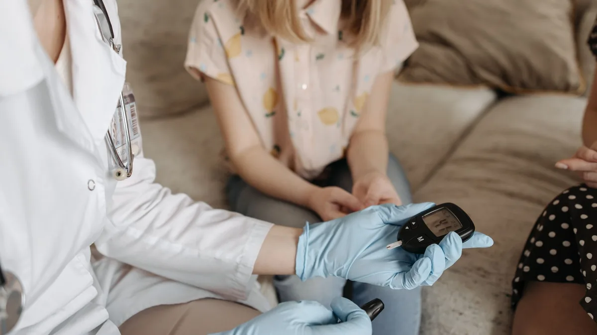
The guideline asks you to check blood sugar at home every day, with some readings 1 to 2 hours after a meal. Check blood pressure daily too. Many people with type 2 diabetes can stop insulin once they need less than 10 units a day. A C-peptide blood test first confirms that your own body still makes insulin. Some people who look like type 2 have a slow form of type 1. Signs include needing insulin within 5 years of diagnosis or not being overweight when diagnosed. They should not stop insulin.
Some nerve-pain medicines are on a separate list in the guideline: drugs that can push sodium too low in the first weeks of low-carb. It names amitriptyline and other tricyclic antidepressants, SNRIs, the seizure drugs carbamazepine and oxcarbazepine, NSAIDs and opioid pain medicines. If you take one, your doctor keeps an eye on your sodium as the carbs come down.
One more note for this site’s readers. Long-term metformin can lower vitamin B12, and low B12 can cause burning feet on its own. See B12 deficiency and burning feet.
Why would my feet burn more after my sugar comes down?
Doctors call it treatment-induced neuropathy of diabetes. It is uncommon, but it is real. A 2024 case report described a 50-year-old man whose A1C was 14.9%. He started a low-carb diet with metformin, and semaglutide was added a week later. His A1C fell to 6.9% in under three months. In the fifth week, both feet began to burn, tighten and go numb. He rated the pain 10 out of 10.
The authors note the risk rises with the size and speed of the A1C drop. The pain can take 12 to 24 months to settle. The guideline makes the same point about the eyes. In people with diabetic eye disease, a fast drop in A1C can briefly make it worse, so that drop should be avoided.
That is why we pace the change. A high starting A1C gets a steady slope, not a cliff.
What should I eat on a low-carb plan for neuropathy?
The guideline defines low-carb as under 130 grams of carbs a day. Its simple list is meat, fish, eggs, dairy and vegetables that grow above the ground. Bread, pasta, rice, cereal, sugary fruit and starchy root vegetables come off the plate. Count total carbs, not “net.” Most people do not need to count calories.
Protein comes first, about 25 to 30 grams per meal. Protein protects muscle, and muscle burns sugar. Fat is eaten to fullness. That fullness is the point. It quiets the cravings that drag people back to sugar.
Dr. Padda adds his own rules for his patients. Industrial seed oils come out. Real olive oil is fine. Red meat and fish are good food. Refined grains stay out. These are his positions. The guideline does not name seed oils.
Kidney disease changes the protein plan. The guideline sets protein near 0.8 grams per kilogram of body weight a day for people with kidney disease. That matters here, because diabetes often hurts the kidneys and the nerves together.
Which blood tests should I ask for before I start?
Dr. Padda learned this the hard way. Early in his career he told patients with prediabetes that their A1C was “not too bad.” He was wrong. Insulin had been climbing for years before that number moved. Now he tests the earlier signals.
The guideline’s baseline is a metabolic panel, a lipid panel, A1C and, with diabetes, a urine protein test. It lists fasting insulin, C-peptide and B12 as options. In our clinic, these belong on the list:
Fasting insulin. Above 10 µIU/mL is our high-risk cutoff.
LP-IR, an insulin resistance score that rises before A1C does.
GGT, an early fatty liver signal. Our cutoff is above 20 U/L.
Oxidized LDL and the omega-6 to omega-3 ratio, which read the processed food in your diet.
Omega-3 index, with a target above 8%.
hs-CRP for inflammation, and 25-OH vitamin D.
B12 with methylmalonic acid, and homocysteine.
One plain note about the guideline itself. It is an expert consensus, built from a panel vote and a non-systematic review of studies. It is not a clinical trial. Dr. Padda holds the society’s Metabolic Health Practitioner credential. He did not write or vote on this guideline.
Where do I start?
Your nerves sit downstream of your blood sugar and your insulin. You can change both, starting with your next meal. Get your baseline labs. Bring your full medicine list to a doctor before day one. Then lower the carbs at a pace your nerves can follow. You are not waiting for the damage to decide your future. You are removing what drives it.
Questions patients ask about low-carb eating and neuropathy
Is it possible to repair diabetic neuropathy?
No treatment reliably regrows nerve. What changes the outlook is removing the driver, so the damage stops adding up and symptoms can ease. Lowering sugar and insulin is one of the strongest moves you can make. See can peripheral neuropathy improve.
Do I need a continuous glucose monitor to start low-carb?
Not always. The guideline cites a study that found a continuous monitor did no better than finger-stick checks. What matters is checking every day and sharing the readings with your doctor, especially in the first week.
Find out what is driving your nerve pain
The free, five-question Nerve Damage Score takes about two minutes and points to the driver most likely behind your symptoms.
Rice SM, Buchanan LA, Calkins MW, Cucuzzella MT, Cywes R, Devine PL, Kalamian M, Kalayjian T, Reynolds DB, Westman EC. A Delphi consensus-based guideline for the implementation of therapeutic carbohydrate reduction in metabolic disorders. Journal of Metabolic Health. 2026;9(1):a152. doi.org
Sahyouni MJ, Acevedo LA, Rodriguez SC, Chiles JA, Pennings NJ. Neuropathy of diabetes following initiation of a low-carbohydrate diet: case report. Obesity Pillars. 2024;11:100115. doi.org
B12 deficiency can cause burning, tingling and numb feet. One of the most overlooked reasons is a stomach that has quietly stopped absorbing the vitamin. If your feet burn and no one has found a cause, ask for more than one B12 level. Ask for methylmalonic acid, both stomach antibodies, iron studies and a medication review.
People with burning feet get sent to look at their spine, their blood sugar or their shoes. Almost nobody gets sent to look at their stomach. Yet the vitamin your longest nerves need cannot enter the body without a protein the stomach lining makes. That chain is the subject of Chapter 6 of The Angry Gut, the new book from Dr. Gurpreet Singh Padda, MD, MBA, MHP, and coauthor Ami Michelle Grimes (announced here).
How can a stomach problem make my feet burn?
Think of B12 as a package that needs an escort. Stomach acid frees it from food. Intrinsic factor, made by the parietal cells of the stomach lining, grabs it and carries it the full length of the small bowel to a receptor at the very end. No escort, no delivery. Only about 1% of the vitamin slips in without that help.1
The nerves that run to your toes are the longest in the body. They sit at the far end of the longest supply line. So they are the first to fail when supply runs short. That is why a B12 problem so often starts in both feet as numbness, pins and needles or burning. Then it climbs toward the calves.
Two things destroy that lining. One is an immune attack on the parietal cells. The other is a long-running H. pylori infection. Either one causes atrophic gastritis. When the autoimmune form causes B12 deficiency, it is called pernicious anemia.
Why was I never told my stomach could be involved?
Because the name points everyone at the wrong test. “Pernicious anemia” suggests a blood-count problem. A 2025 perspective on the English national guideline notes that anemia is present in only 15% to 20% of pernicious anemia diagnoses.2
In the chapter, I walked past the four years before this diagnosis. Those were the years when the lab work kept coming back fine. That was the wrong place to stop. The stomach fails in an order, and B12 is near the end of it.
Iron usually goes first. Sometimes the damage sits in the upper, acid-making part of the stomach. Then up to half of patients run low on iron, often well before B12 drops.3
It is rarely one nutrient. One center tracked 122 patients diagnosed by biopsy. Of those, 76 were short on at least one of B12, iron, vitamin D or folate, and 52 were short on more than one.4
The wait is long. The median delay to diagnosis in the autoimmune gastritis literature is 14 months.4
The lining does not wait with you. A prospective study followed 270 patients. The stomach tissue of every one of them got worse over time, usually within three years. None got better on its own.4
Who is most likely to have a stomach-driven B12 problem?
The people already at risk for nerve damage carry the highest odds.
Type 1 diabetes. The risk of autoimmune gastritis runs about three to five times that of the general public.4
Thyroid disease. Autoimmune thyroiditis shows up in 36% to 44% of people with autoimmune gastritis. Turned around, about 40% of people with autoimmune thyroid disease have this kind of stomach atrophy.4 If you have Hashimoto’s and burning feet, both belong in the same talk. See how thyroid function affects your nerves.
Long-term metformin. In the Diabetes Prevention Program, adults at high risk for type 2 diabetes were randomized to metformin (1,073 people) or placebo (1,082). At five years, low or borderline-low B12 (at or below 298 pg/mL) showed up in 19.1% on metformin. On placebo it was 9.5%. The extra neuropathy on metformin showed up only in the people whose B12 had fallen low.5 Never stop metformin on your own. Ask for the B12 workup instead. More in how statins and metformin can affect nerve health.
Daily acid blockers. A large study compared patients with and without B12 deficiency. People taking more than 1.5 proton pump inhibitor pills a day had nearly double the odds of B12 deficiency (odds ratio 1.95).6
Weight-loss surgery. A Portuguese hospital tracked 505 patients in a row. At twelve months, B12 deficiency ran 16.74% after gastric bypass. After sleeve gastrectomy it was 0.93%.7 See nerve damage after stomach or bowel surgery.
Follow the money and the list stops looking random. We subsidize cheap calories. The calories cause the diabetes. The diabetes earns the metformin. And the metformin drains the vitamin while one blood test keeps saying all is fine. None of that is the patient’s failure.
What should I have measured if my feet burn?
A single B12 level tells you how much vitamin is in the blood. It does not tell you whether your nerves are getting it (see why a normal B12 can miss a deficiency). Bring this fuller list to your visit.
Serum B12, read against the right lines. In American primary care, below 180 pg/mL is diagnostic and 180 to 350 pg/mL is borderline.8
Methylmalonic acid if you land in that borderline band. It rises when B12 fails inside the cell. Kidney problems, dehydration, low thyroid and bacterial overgrowth in the small bowel can also raise it. So it has to be read with those in mind.9
Both stomach antibodies, together. Anti-parietal cell and anti-intrinsic factor antibodies each miss cases on their own. Run as a pair, they reached 90% sensitivity and 95.7% specificity in a 2025 comparison of assays in autoimmune gastritis.10 A negative intrinsic factor antibody alone does not rule the problem out. It tends to turn positive later in the disease.3
Ferritin and iron studies, including the oldest ones in your chart. A low iron years ago may have been the first clue.
A medication history that counts years of metformin and daily dose, and years of acid blockers.
Gastrin only after an acid blocker is stopped. A proton pump inhibitor can raise gastrin three to five times. That makes the result useless.2
Do not wait for large red blood cells either. They are a late sign, and other problems can cause them too. Some patients low on B12 show nothing wrong on the blood count at all.9
Does it change treatment if my stomach is the cause?
Yes. When the absorption machinery is gone, a supplement bottle is a gamble on that 1% side door. England’s 2024 national guideline offers lifelong B12 shots when autoimmune gastritis is the cause or suspected cause, or after total gastrectomy.11 In the chapter’s case, a man whose stomach had almost no parietal cells left went on shots, not tablets. Eleven months later his walking was steadier. Some of what he lost did not come back. That is the cost of finding it late.
A burning foot is a symptom. The cause may be hiding in the stomach, the medication list or the iron history. You can ask about all of them this week. You do not have to accept “idiopathic” while those questions are still open. The chapter’s free technical supplement is at theangrygut.com/chapter-6.
Questions patients ask about B12 and burning feet
Can low B12 cause burning feet even if I am not anemic?
Yes. Anemia shows up in only a minority of pernicious anemia diagnoses. So a normal blood count does not rule out a B12 problem reaching the nerves. See what causes burning, numb or painful feet.
I have diabetes. How do I know if it is B12 and not my blood sugar?
You often cannot tell from symptoms alone, and both can be present. Metformin use, type 1 diabetes and thyroid disease all raise the odds of a B12 problem. That is why a diabetic patient with burning feet deserves B12 testing too. See diabetic peripheral neuropathy treatment in St. Louis.
Should I just start taking a B12 supplement?
Get tested first. If the stomach can no longer absorb the vitamin, a tablet depends on that 1% side door. England’s national guideline recommends shots instead when autoimmune gastritis is the cause. The form and dose are a medical decision. See nutritional deficiencies beyond B12.
Sources
Chan CQH, Low LL, Lee KH. Oral vitamin B12 replacement for the treatment of pernicious anemia. Frontiers in Medicine. 2016;3:38. doi.org
Thain A, Hart K, Ahmadi KR. Addressing the gaps in the vitamin B12 deficiency 2024 NICE guidelines: highlighting the need for better recognition, diagnosis, and management of pernicious anaemia. European Journal of Clinical Nutrition. 2025;79(7):607–610. doi.org
Shah SC, Piazuelo MB, Kuipers EJ, Li D. AGA clinical practice update on the diagnosis and management of atrophic gastritis: expert review. Gastroenterology. 2021;161(4):1325–1332. doi.org
Rustgi SD, Bijlani P, Shah SC. Autoimmune gastritis, with or without pernicious anemia: epidemiology, risk factors, and clinical management. Therapeutic Advances in Gastroenterology. 2021;14:17562848211038771. doi.org
Aroda VR, Edelstein SL, Goldberg RB, et al. Long-term metformin use and vitamin B12 deficiency in the Diabetes Prevention Program Outcomes Study. Journal of Clinical Endocrinology and Metabolism. 2016;101(4):1754–1761. doi.org
Lam JR, Schneider JL, Zhao W, Corley DA. Proton pump inhibitor and histamine 2 receptor antagonist use and vitamin B12 deficiency. JAMA. 2013;310(22):2435–2442. doi.org
Vieira de Sousa JP, Santos-Sousa H, Vieira S, et al. Assessing nutritional deficiencies in bariatric surgery patients: a comparative study of Roux-en-Y gastric bypass versus sleeve gastrectomy. Journal of Personalized Medicine. 2024;14(6):650. doi.org
Patel H, McGuirk R. Vitamin B12 deficiency: common questions and answers. American Family Physician. 2025;112(3):294–300. pubmed.ncbi.nlm.nih.gov
Harrington DJ, Stevenson E, Sobczyńska-Malefora A. The application and interpretation of laboratory biomarkers for the evaluation of vitamin B12 status. Annals of Clinical Biochemistry. 2024;62(1):22–33. doi.org
Osmola M, Hémont C, Romańczyk M, et al. A comparative study of different assays for autoantibodies detection in patients with autoimmune gastritis. Journal of Translational Autoimmunity. 2025;10:100294. doi.org
National Institute for Health and Care Excellence. Vitamin B12 deficiency in over 16s: diagnosis and management (NG239). 2024. nice.org.uk
Find out what is driving your burning feet
The free, five-question Nerve Damage Score takes about two minutes and points to the driver most likely behind your symptoms.
Whether your neuropathy gets worse depends far more on one thing than on the word “neuropathy” itself. Is the driver damaging your nerves still active? That is the whole answer. It is also why nobody can give you a timeline until the driver has been found.
Peripheral neuropathy is the most common reason for an outpatient neurology visit in the United States. It accounts for over $10 billion in health care spending each year.1 Much of that reflects how long people live with a diagnosis that names the symptom, not the cause. Progression is not one fixed path, and it can be measured.
How does neuropathy progress?
When people say progression, they usually mean symptoms. But symptoms are only one of the things that change. Feeling, balance, strength and daily function can all shift, sometimes in different directions at once.
Length-dependent and symmetrical patterns
In many metabolic forms, the longest nerve fibers fail first. That is why symptoms usually start in the toes and move upward. It is also why the hands are often affected later, once the pattern has reached about mid-calf.
The pattern is also usually the same on both sides. That points toward a whole-body (systemic) driver. A one-sided or patchy pattern points somewhere else entirely. That is a useful thing for your clinician to notice early.
Small fiber involvement can progress when standard testing looks normal
Burning, hot-coal feelings and changed pain and temperature sense can be present while a routine nerve conduction study looks normal. That is because the study measures large fibers.
A normal result in that setting does not mean nothing is getting worse. It is a signal that the workup needs to match the symptom pattern, not stop.
What causes neuropathy to get worse?
Peripheral neuropathy is a category, not a diagnosis. These are the driver groups worth working through. More than one can be present in the same person.
Metabolic injury
When glucose runs high, sugars stick to proteins in a slow browning reaction. That makes advanced glycation end-products, which build up around the nerve. Poor flow through the small vessels that feed the nerve makes it worse. That is because the longest fibers depend on the weakest part of that supply.
The practical point: while the metabolic driver runs, injury goes on. That is true whether or not symptoms happen to be quiet that week. How strong the symptoms feel and how active the disease is are two different things.
Nutritional drivers
B12 deficiency can cause nerve damage that keeps getting worse. It keeps going until the deficiency is corrected. Diet, absorption after gut surgery, and drugs that interfere with absorption all belong in the history.
Autoimmune and immune-reactive patterns
Immune-driven processes can keep symptoms advancing while the immune activity goes on. Gluten-related nerve injury is one of them. These patterns are often the ones hiding behind an idiopathic (no known cause) label.
Toxic and medication-related exposures
Chemotherapy-related nerve injury follows the timeline of the exposure. So the history and the course of symptoms have to be read together. Heavy metals such as lead, arsenic and mercury are the other exposure group. They are worth ruling in or out when the history suggests it.
Structural compression
Not everything that looks like neuropathy is systemic. Sometimes symptoms follow the path of a single nerve. Then progression may be driven by pressure on the nerve (compression) and local inflammation instead. That responds to entirely different treatment. The difference is covered in nerve compression: carpal tunnel, sciatica and double-crush patterns.
How is neuropathy progression measured?
Progression is change over time. Change over time only means something against a baseline. Without one, every visit is a fresh guess about whether things are moving.
We perform electrodiagnostic testing (EMG/NCS) on site. It measures how large fibers carry signals. When unsteadiness, dizziness or falls are part of the picture, we use VNG as a diagnostic test of inner-ear balance function. It is not a treatment, and it does not measure peripheral nerves.
Metabolic and nutritional testing runs alongside both, because that is where most of the drivers you can change actually live.
Can neuropathy damage be reversed?
We do not promise that nerve damage can be regenerated or reversed. A plan aimed at the driver has a different goal, and one that is easier to defend. It finds what is still causing injury. It reduces that injury where it can be reduced. And it protects function.
Symptom treatment has a place inside that. Qutenza (capsaicin 8% patch) is FDA-approved in adults only for neuropathic pain from postherpetic neuralgia and from diabetic peripheral neuropathy of the feet. It is applied in clinic by a clinician and repeated no more often than every three months.
Orthobiologic injections such as PRP and BMAC are offered in selected situations. Better sleep and less pain are worth having in their own right. But neither one changes the driver.
When should worsening neuropathy be checked right away?
Some changes are worth acting on quickly instead of watching.
Numbness that is spreading upward, or newly involving the hands.
Pain that has started to disrupt sleep, or pain set off by light touch such as a bedsheet.
New unsteadiness, dizziness, or any fall.
Difficulty with stairs, weakness, or a foot that catches when you walk.
Any break in the skin, blister, or hot, swollen foot when feeling is reduced.
A high-risk history: diabetes, chemotherapy, known B12 risk, or a major exposure.
When protective feeling is reduced, checking your feet every day stops being optional. A numb foot no longer reports injury. So your eyes have to do the job the nerves used to do.
Start with the Nerve Damage Score
The Nerve Damage Score is five questions, and it is free. It is built to point at the driver most likely responsible in your case. That is the only honest starting point for a talk about whether things will get worse.
Regenerve, 4477 Woodson Rd #104, St. Louis, MO 63134. Call (314) 886-5739 or text (314) 886-5902, or email info@regenerve.com. We are minutes from St. Louis Lambert International Airport and see patients from across the St. Louis region, Missouri and Illinois, including Edwardsville, Glen Carbon, Collinsville, Troy and Maryville.
Frequently asked questions
My feet burn at night. Does that mean it is getting worse?
Not by itself. Nighttime burning is common early and does not reliably track severity. So how bad it is on a given night is a poor measure of progression. What matters is whether feeling, balance and function are changing over months. See small fiber neuropathy symptoms and testing.
How do clinicians predict whether neuropathy will progress?
By finding the driver and then watching whether it is still active. A driver that is being corrected and a driver that is left alone lead to very different paths in the same person. See the hidden drivers of peripheral neuropathy.
My blood sugar is under control now. Am I safe?
Better glucose control reduces one source of ongoing injury, but it is not the whole terrain. Lipids, blood pressure, small blood vessel health, nutrition status and any other driver all still matter. See diabetic peripheral neuropathy treatment in St. Louis.
What if nobody has found a cause for my neuropathy?
Idiopathic is a status, not a verdict. It usually means the workup stopped too soon, before the less common drivers were looked at. These include deficiency states, immune-reactive patterns and exposures. See functional B12 deficiency and neuropathy.
Could an exposure be making mine worse without my knowing?
It is possible, and it is worth ruling in or out instead of assuming. Heavy metals such as lead, arsenic and mercury are the classic example. Exposure history is often the only clue before testing. See heavy metals and neuropathy.
How do you stop neuropathy from progressing?
Find the driver that is still causing injury and reduce it where it can be reduced. That can mean correcting a B12 deficiency, getting metabolic injury under control, or removing a toxic exposure. A baseline from nerve testing lets your clinician see whether things are actually changing. Symptom treatment helps you feel better, but it does not change the driver.
Can neuropathy spread to the hands?
Yes. In many metabolic forms the longest nerve fibers fail first, so symptoms usually start in the toes and move upward. The hands are often affected later, once the pattern has reached about mid-calf. Numbness that newly involves the hands is one of the changes worth getting checked promptly instead of watched.
What’s the worst that can happen with neuropathy?
When protective feeling fades, a numb foot stops reporting injury. So a blister, a break in the skin or a hot, swollen foot can go unnoticed. Weakness, a foot that catches when you walk, new unsteadiness and falls are the other serious turns. Each one needs a prompt check. Daily foot checks become essential once feeling is reduced.
Sources
Tavee JO. Office approach to small fiber neuropathy. Cleveland Clinic Journal of Medicine. 2018;85(10):801–812. ccjm.org
Whole-body infrared delivers infrared light and radiant heat to the entire body, not to one limb or one foot. At Regenerve it is supportive care. It sits inside a plan built around the specific driver of your neuropathy — metabolic, nutritional, autoimmune, toxic or structural. It is never a treatment given on its own.
Two things are true at once, and this page holds both. Medicare does not cover infrared therapy devices for peripheral sensory neuropathy, so you pay for a course out of pocket.2 And the randomized evidence behind that determination studied a different delivery method. It used narrowly selected patients, for a few weeks at a time. Neither fact settles what infrared can do for one specific person. This page explains why.
Does Medicare cover infrared therapy for neuropathy?
Effective October 24, 2006, the Centers for Medicare & Medicaid Services made a ruling on infrared therapy devices and related accessories. It found them not reasonable and necessary under section 1862(a)(1)(A) of the Social Security Act. Infrared and near-infrared light and heat are not covered anywhere in the country for diabetic and non-diabetic peripheral sensory neuropathy. That includes monochromatic infrared energy. The same goes for wounds and ulcers of the skin and the tissue under it (subcutaneous tissues). It also goes for symptoms such as pain from those conditions. The policy is on record as national coverage determination 270.6. It was implemented January 16, 2007.2
Know this before you start anything, because it decides how the service is paid for. Patients sign an advance beneficiary notice for non-covered services. Then they pay out of pocket. Anyone who offers you infrared for neuropathy and implies it will be billed to Medicare is either mistaken or misleading you.
A coverage determination is not a clinical finding about your nerves. It is a program’s decision about what it will buy. It was made under a legal standard, at a fixed point in time, on the evidence available then. That is a real and important decision. But it answers a different question from whether a therapy helps a particular person with a particular driver. The rest of this page deals with that clinical question.
What is the difference between infrared pads and whole-body infrared?
The word covers a wide band, and the difference is not cosmetic. It decides which studies apply to what you are being offered.
Contact pad devices
These are rows of small infrared lights (diodes) set in flexible pads. The pads are strapped against the skin of the foot and lower leg. This is the device the 2006 coverage determination describes and regulates. It falls under the durable medical equipment benefit category.2 With very few exceptions, it is also what the randomized neuropathy studies tested. In this field the technique is usually called monochromatic infrared energy.
Whole-body radiant infrared
This is a cabin or enclosure that raises body temperature with radiant infrared. There is no pad. There is no fixed wavelength aimed at a target tissue. The dose is very different, and so is the amount of tissue it reaches. In regulatory terms, infrared lamps emit across roughly 700 to 50,000 nanometers and are meant to provide topical heating (heating at the skin).3
When you compare what is on offer, ask which of these two you are actually buying. A study of pads at 890 nanometers on the sole of a foot does not automatically describe a warm cabin, in either direction.
What do the infrared therapy trials for neuropathy show?
A 2017 systematic review and meta-analysis in the Brazilian Journal of Physical Therapy pooled six randomized controlled trials. They covered 594 feet in 304 patients with diabetic sensorimotor polyneuropathy (nerve damage from diabetes).4 It is the best single place to see this body of work. Its own rules set the limits of what it can tell you.
The review included randomized trials of light-emitting diodes in the 750 to 1300 nanometer range. By rule, it left out any trial that applied the treatment to a region other than the feet. It also left out any trial with fewer than three days of treatment.4 Every device in it was a pad set placed on the foot and lower leg, at 870 to 890 nanometers. Each delivered 1.30 to 1.95 joules per square centimeter per minute. Follow-up ran from one and a half to twelve weeks, with a median of four weeks.4
The pooled result
Across four trials and 402 feet, infrared did not increase the number of plantar regions (spots on the sole) that could feel the 5.07 monofilament, compared with control. The standardized mean difference was 0.22, 95% confidence interval −0.07 to 0.51.4 A subgroup with follow-up of two weeks or less did show a gain: standardized mean difference 0.41, 95% confidence interval 0.18 to 0.64. That gain was gone beyond two weeks: standardized mean difference −0.12, 95% confidence interval −0.50 to 0.26. The difference between those subgroups was itself statistically significant at p = 0.02.4
For pain, the review set aside one non-blinded trial. After that, pain fell less in the infrared groups than in the sham groups: mean difference 0.40 points, 95% confidence interval 0.30 to 0.68.4 The review’s authors offer a specific reading of that, and it is worth knowing. In a nerve that is losing fibers, restoring some feeling can restore the ability to feel pain. So falling pain scores in this disease are not always good news.4 Using the GRADE system, the authors rated the overall certainty of evidence as low for both outcomes. That means further studies are likely to change the estimate.4
The largest and longest of the trials
A 2008 double-blind, sham-controlled trial in Diabetes Care randomized 69 people with diabetes. Of those, 60 patients and 120 limbs completed it. Units were used at home for 40 minutes a day for 90 days. The investigators measured nerve conduction velocities and vibration perception threshold. They used four grades of Semmes-Weinstein monofilament. They used the Michigan Neuropathy Screening Instrument. They also used a 10 cm visual analog pain scale and a quality of life tool built for neuropathy. There were no significant differences between active and sham on any of them.5
The entry rule that matters here is spelled out in the published report. Entry required a vibration perception threshold between 20 and 45 volts.5 That is a severity band. People with milder loss below that band were not enrolled. Neither were people with the most profound loss above it. We could not obtain the full eligibility list from the publisher. So we do not describe any exclusions beyond the one the report states.
The trial where both groups improved
A 2005 randomized, double-blind, placebo-controlled study in Diabetes Care followed 39 people. All had diabetic peripheral neuropathy. They got 30 minutes of active or placebo infrared three times a week for four weeks. Then they had four weeks with no treatment. The number of sites that could feel the 5.07 monofilament rose significantly in both the active and the placebo groups. There were no significant differences between the groups at any measurement point.6
The Medicare review of this trial recorded the size of that placebo movement. Over the blinded phase it was about +0.37 sites in the active arm and +0.57 sites in the sham arm, roughly 39% and 40% above baseline.3 This is the single most useful thing in the whole literature. It is why a clinic showing you before-and-after numbers with no control group has shown you nothing. And it applies to glowing reports about any modality, not only this one.
The trial that separated symptoms from structure
A 2021 randomized sham-controlled study in Neurology India enrolled 38 people with type 2 diabetes and painful diabetic peripheral neuropathy. Of those, 30 completed it. Treatment was three times a week for 12 weeks. Intraepidermal nerve fiber density (the count of tiny nerve endings in the skin) was measured on a 3 mm skin punch biopsy at baseline and at 12 weeks. Mean age was 59.1 years. Mean diabetes duration was 12.9 years, and mean symptom duration was 3.9 months.7
Nerve fiber density did not increase with treatment. Pain did move. The median visual analog scale score fell 5.1 points with infrared against 3.0 points with sham, intergroup p = 0.01. Quality of life on the Norfolk instrument also differed, at p = 0.021. The authors concluded that infrared does not increase nerve fiber density over short-term use. But they found it does provide symptom benefit and improved quality of life.7
For a patient deciding what to expect, this is the most informative single trial. It is also the clearest example of a narrow population. Mean symptom duration was under four months. People with years of symptoms were not in it. Neither were people with type 1 diabetes, or people whose neuropathy is not diabetic at all.
The smallest of the randomized trials
A 2011 single-blinded randomized controlled trial in the Singapore Medical Journal studied 30 feet from 24 patients. They were randomized to 12 daily infrared or sham treatments. Current perception threshold was measured by neurometer at 2,000 Hz, 250 Hz and 5 Hz, and checked again at six weeks and three months. No significant difference was found between the infrared and sham feet.8 The published report does not state a full exclusion list that we could obtain.
The other side of the argument
A 2023 systematic review in Current Diabetes Reviews took a wider view of photobiomodulation (light therapy aimed at cells) in diabetic peripheral neuropathy. It covered eight studies in people with type 2 diabetes. It reported less nerve pain (neuropathic pain) and better nerve conduction velocity. It also reported lower plantar pressure distribution (pressure across the sole). Its authors concluded that photobiomodulation is an effective, non-invasive option in this condition.9
That conclusion is real, and it belongs on this page. So does its basis. The review deliberately included both randomized and non-randomized studies,9 and uncontrolled designs are exactly where the placebo movement described above does its damage. It is a narrative synthesis, not a pooled estimate. It is limited to type 2 diabetes. A reader has every right to weigh it. A reader has no right to be told it settles the question.
Two independent groups have registered protocols for new systematic reviews in this area. One was published in BMJ Open in 2022 and one in PLOS ONE in 2024. Both call for randomized trials, with pain as a primary outcome. Their secondary outcomes include nerve conduction, sensation, quality of life and adverse events.1,10 Protocols describe what a good answer would look like. They are not answers themselves. But two of them, registered fifteen years after a coverage determination, are a fair signal. Researchers consider the question open, not closed.
What the 2006 decision was actually based on
Coverage decisions are documents, and this one is public. Reading it is more useful than repeating its conclusion.
Before the analysis there was no national ruling on these devices. But local Medicare contractors for durable medical equipment already had the same non-coverage rules. In the peripheral neuropathy literature, the analysis found 14 studies. Of those, 11 had an infrared-only arm. Only three of those were placebo-controlled with a parallel group run at the same time. All three were double-blinded and used sham devices. They enrolled 18, 43 and 50 patients.3
The blinded treatment periods in those three trials were two weeks in one and four weeks in the other two. Between them they tested only two wavelengths, 890 and 905 nanometers. No investigator did dose-ranging (testing a range of doses) for energy density, dosing frequency or dosing length. None varied pulsatility or duty cycle. None checked whether any effect lasted long-term. None used hard clinical endpoints such as ulcer or amputation rates.3 The analysis stated that despite an exhaustive search it found no high-quality randomized phase III trials with hard clinical endpoints. It also stated that a major placebo effect ruled out confident reading of the many studies in which patients served as their own controls.3 It noted that two large trials were then in progress. They could not be reviewed because they were not yet complete or published.3
That is a defensible decision on that record. It is also a decision about three small, short, single-wavelength trials of a contact pad device.
What has changed since
Four of the six trials in the 2017 meta-analysis were published after the determination took effect.4 Trial lengths grew from four weeks to 90 days5 and to 12 weeks.7 Outcome measures widened from monofilament counts to nerve conduction velocity and quality of life.5 Then they widened to intraepidermal nerve fiber density on skin biopsy.7
The overall picture did not reverse. The pooled estimate for sensation is still null, with low certainty. The pain estimate does not favor infrared.4 The real additions on the other side are two. One is a symptom and quality of life signal without a structural change.7 The other is a narrative review reporting benefit from a mixed body of randomized and non-randomized studies.9
What has not changed
The coverage policy itself. The public record for national coverage determination 270.6 shows coding updates for ICD-10 in 2015 and 2017. The policy did not change. It states that the determination was last reviewed in November 2006.2 Anyone who claims Medicare recently shifted its view on infrared for neuropathy is describing something that is not in the record.
Three research gaps also remain exactly where they were. There is still no phase III trial with hard clinical endpoints. There is still no dose-ranging study (one that tests a range of doses). No study has set what energy density, session length or course length would even be the right thing to test. And there is still no randomized trial of whole-body delivery in peripheral neuropathy at all — the pooled review excluded use on any region other than the feet, under its own eligibility rules.4
That last point needs saying carefully, because it is easy to abuse in both directions. A lack of trials of whole-body infrared is not evidence that whole-body infrared works. It is also not what the null pad-device results measured. It is a gap. A gap is a reason for modest expectations and honest conversation. It is not a reason for confidence either way.
The patients these trials did not study
Randomized trials earn their authority by controlling variation. They control it by choosing who gets in. That choice shows in this literature wherever the reports state it.
A severity band. The 90-day home-use trial required a vibration perception threshold between 20 and 45 volts.5 Milder and more profound loss were both outside it.
A region rule. The 2017 meta-analysis left out any trial that applied the treatment outside the feet, and any trial shorter than three days.4
An early-symptom population. The nerve fiber density trial enrolled people with a mean symptom duration of 3.9 months. Their mean diabetes duration was 12.9 years, and all had type 2 diabetes.7
One form of diabetes. The 2023 light therapy review (photobiomodulation) was limited to type 2 diabetes.9
For several of these trials we could not obtain the full eligibility and exclusion list from the publisher. In those cases we have not guessed at exclusions. This page does not claim any rule a report does not state.
What we can verify is what the designs did not test. None of these trials studied infrared in someone whose insulin resistance was being actively treated. None studied someone whose B12 deficiency had just been corrected, whose autoimmune driver had been found, or whose toxic exposure had been removed. None enrolled a person carrying several drivers at once alongside a long medication list. And every one of them tested infrared alone against a sham, on a fixed schedule, for a few weeks.
So a null result has limits. It came from a selected population, for one modality used by itself, over a short period. It tells you this device, at these settings, in these people, over this interval, did not beat sham on these measures. It does not show what happens in the patients who were screened out. And it does not describe a therapy used as one part of a plan instead of as the plan. That is the ordinary gap between efficacy under trial conditions and effectiveness in practice.
It cuts both ways, and we say so. The same logic that stops a null trial from closing the question also stops it from being a license to assume benefit. Where nobody has measured, nobody knows.
Does infrared heat improve circulation to the nerves?
There is a real reason clinicians care about heat and blood flow. It is separate from the neuropathy trials above.
What the heat therapy evidence shows
A 2021 systematic review and meta-analysis in Experimental Physiology pooled 15 studies of heat therapy. They covered healthy people and patients. Exposures ran 30 to 90 minutes over 10 to 36 sessions. Compared with control, heat therapy cut mean arterial pressure (average blood pressure) by 5.86 mmHg. It cut systolic blood pressure by 3.94 mmHg and diastolic blood pressure by 3.88 mmHg. It improved brachial artery flow-mediated dilatation (how well the arm artery widens) by 1.95%. Resting heart rate was unchanged. Certainty was rated moderate for the blood pressure outcomes. It was rated low for mean arterial pressure and flow-mediated dilatation.11
Those are heart and blood vessel outcomes, from a body of work about heat therapy in general. Not one of them is a nerve measurement. They give a reasonable mechanistic reason to be interested in whole-body heat when neuropathy has a microvascular (small blood vessel) component. They are not evidence of nerve benefit. We do not present them that way, and neither should anyone else.
The vasa nervorum question
Peripheral nerves are fed by very small vessels, the vasa nervorum. The longest nerves in the body depend on the most fragile end of that supply. Oxygen delivery is a real bottleneck in metabolic neuropathy. That is why treatments that change local blood flow attract attention.
Warmth causes vasodilation (widening of blood vessels). That much is not in dispute. Whether raising whole-body temperature on a schedule produces a lasting change in nerve function is a different claim. It has not been shown in a randomized trial in this condition.
Metabolic stress
Infrared is sometimes described as modulating metabolic stress in nerve tissue. We have not found trial evidence testing that claim in people with peripheral neuropathy. So we do not make it. And we do not use it as a reason to delay the metabolic and nutritional work that does have evidence behind it.
Is infrared heat therapy safe if you have neuropathy?
This is not a theoretical concern and it is specific to heat.
Loss of feeling in diabetic peripheral neuropathy raises the risk of burns, injuries and foot ulceration (foot sores).12 The mechanism is simple. The warning system that tells you something is too hot is the same system the disease has damaged. In the infrared studies themselves, the Medicare review noted that two patients in one trial got surface burns (superficial burns) under the device pads.3
That is why heat-based therapy in neuropathy belongs under clinical supervision. Skin is inspected before and after sessions. Session settings are set by someone who has measured how much sensation you have lost. And there is a low bar for stopping. It is also why a home device bought online carries a different risk from a supervised session, when the person using it cannot feel a burn forming.
Where whole-body infrared sits in a plan at Regenerve
Regenerve is directed by Dr. Gurpreet Singh Padda, MD, MBA, MHP. Whole-body infrared is one of the modalities available here. Others are class 4 photobiomodulation, class 3B cold laser, and metabolic and nutritional care. Electrodiagnostic testing (EMG/NCS) is done on site. VNG is a diagnostic test of inner-ear balance function, used when unsteadiness is part of the picture. It is a test, not a treatment.
None of it is prescribed before the driver is mapped. Peripheral neuropathy is a category, not a diagnosis. The order of steps matters more than the equipment list — a point covered in the hidden drivers of peripheral neuropathy.
What we tell patients about infrared specifically
We tell them the coverage status, so there is no billing surprise. We tell them the trial evidence is for a pad device on the feet. It is null on sensation over the lengths of time studied. The one trial measuring nerve fiber density found no structural change, while reporting symptom and quality of life benefit. We tell them no randomized trial has studied the whole-body form at all. We describe what we expect it to add — comfort, warmth, tolerance for the activity and metabolic work that carry the plan. And we say plainly that we are not claiming it repairs nerves.
We set a review point before starting. We agree on what would count as a change, and we stop if nothing changes. We do not promise that nerve damage can be regenerated or reversed. We publish no success rate for any Regenerve protocol.
“My ethos is to treat all of my patients as I would my own family, with the goal of giving them back their quality of life.” — Dr. Gurpreet Singh Padda, MD, MBA, MHP
Questions worth asking before starting a course
Is this a contact pad device at a stated wavelength and dose, or whole-body radiant heat? The published trials are about the first one.
How was the driver of my neuropathy found before this was recommended, and what tests were used?
What outcome will be measured, with what instrument, and at what interval?
How is my reduced ability to feel heat being accounted for in the protocol, and who inspects my skin?
Is this being offered as one part of a plan, or as the plan?
What is the review point, and at what stage do we stop?
Am I signing an advance beneficiary notice, and what will I owe?
Start with the Nerve Damage Score
The Nerve Damage Score is five questions, and it is free. It points to which driver to chase first. That is the only thing that makes any decision about infrared meaningful.
Regenerve, 4477 Woodson Rd #104, St. Louis, MO 63134. Call (314) 886-5739 or text (314) 886-5902, or email info@regenerve.com. We are minutes from St. Louis Lambert International Airport and see patients from across the St. Louis region, Missouri and Illinois, including Edwardsville, Glen Carbon, Collinsville, Troy and Maryville.
Is whole-body infrared therapy proven to treat peripheral neuropathy?
Not in the sense of a proven treatment. The randomized trials in this area studied contact pad devices on the feet and lower legs. A 2017 meta-analysis of six of them found a short-term gain in monofilament sensation that did not last, on low-certainty evidence. No randomized trial has tested whole-body delivery in peripheral neuropathy. So for that specific form there is an evidence gap, not a negative finding. Since 2006, Medicare has not covered infrared therapy devices for peripheral sensory neuropathy. So you pay out of pocket. We use it as supportive care inside a driver-directed plan, never as the treatment. See what we actually provide on site.
Does infrared help if my neuropathy is diabetic?
Diabetic peripheral neuropathy is driven by metabolic and microvascular (small blood vessel) injury. So the work that changes its course is metabolic. One 12-week sham-controlled trial in painful diabetic neuropathy reported symptom and quality of life benefit. It found no increase in nerve fiber density. That is a fair picture of what to expect: infrared may make you more comfortable while the metabolic work does the structural job. It does not replace that work. See diabetic peripheral neuropathy treatment in St. Louis.
Will infrared help if my neuropathy comes from a B12 deficiency?
It will not correct the deficiency, and correcting the deficiency is the point. Heat and light do nothing about absorption, intake, or the medications that interfere with B12. Infrared can sit alongside that correction for comfort. It cannot replace it. See functional B12 deficiency and neuropathy.
What about gluten-related neuropathy?
Same answer, different mechanism. If an immune reaction to gluten is driving the nerve injury, the treatment that matters is dietary and immune-based, not heat. Also note that people with non-diabetic drivers were not enrolled in the infrared trials at all. So there is no trial evidence either way for this group. See the gluten and neuropathy connection.
My nerve conduction study was normal but my feet still burn. Would infrared help?
A normal conduction study with ongoing burning points toward small fiber involvement. Standard nerve conduction testing often misses that. The next step is a workup matched to that pattern, not another modality, because a modality chosen before the driver is found is a guess. See small fiber neuropathy symptoms and testing.
Can infrared therapy reverse nerve damage?
We do not claim it can. The one trial that measured nerve fiber density found no structural change, even though patients reported better symptoms and quality of life. We use whole-body infrared for comfort, warmth and tolerance for the activity and metabolic work that carry the plan. We do not promise that nerve damage can be regenerated or reversed.
Is it safe to use a home infrared device for neuropathy?
It carries a different risk from a supervised session. Neuropathy damages the same warning system that tells you something is too hot, so a burn can form without being felt. In one infrared trial, two patients got superficial burns under the device pads. Under supervision, skin is inspected before and after sessions and settings match how much sensation you have lost.
Is whole-body infrared the same as infrared pads or laser therapy?
No. Whole-body infrared is a cabin that raises body temperature with radiant heat. Contact pad devices strap infrared diodes against the foot and lower leg. Those pads are what most neuropathy trials studied. Class 4 photobiomodulation and class 3B cold laser are separate modalities offered at Regenerve. Ask which one you are being offered, because studies of one do not describe the others.
Sources
Wang JY, Huang ZQ, Deng HP, Zhao L, Deng HY, Liu JP, Shen XY, Cheng K. Low level light therapy/photobiomodulation for diabetic peripheral neuropathy: protocol of a systematic review and meta-analysis. BMJ Open. 2022;12(9):e059476. pmc.ncbi.nlm.nih.gov
Centers for Medicare & Medicaid Services. National Coverage Determination 270.6, Infrared Therapy Devices. Effective October 24, 2006; implemented January 16, 2007. Medicare Coverage Database. cms.gov
Centers for Medicare & Medicaid Services. Decision Memorandum for Infrared Therapy Devices (CAG-00291N). Medicare Coverage Database. October 24, 2006. cms.gov
Robinson CC, Klahr PDS, Stein C, Falavigna M, Sbruzzi G, Plentz RDM. Effects of monochromatic infrared phototherapy in patients with diabetic peripheral neuropathy: a systematic review and meta-analysis of randomized controlled trials. Brazilian Journal of Physical Therapy. 2017;21(4):233-243. pmc.ncbi.nlm.nih.gov
Lavery LA, Murdoch DP, Williams J, Lavery DC. Does anodyne light therapy improve peripheral neuropathy in diabetes? A double-blind, sham-controlled, randomized trial to evaluate monochromatic infrared photoenergy. Diabetes Care. 2008;31(2):316-321. pubmed.ncbi.nlm.nih.gov
Clifft JK, Kasser RJ, Newton TS, Bush AJ. The effect of monochromatic infrared energy on sensation in patients with diabetic peripheral neuropathy: a double-blind, placebo-controlled study. Diabetes Care. 2005;28(12):2896-2900. pubmed.ncbi.nlm.nih.gov
Rastogi A, Uppula P, Saikia U, Bhansali A. Effect of monochromatic infrared energy on quality of life and intraepidermal nerve fiber density in painful diabetic neuropathy: a randomized, sham control study. Neurology India. 2021;69(5):1331-1337. pubmed.ncbi.nlm.nih.gov
Nawfar SA, Yacob NB. Effects of monochromatic infrared energy therapy on diabetic feet with peripheral sensory neuropathy: a randomised controlled trial. Singapore Medical Journal. 2011;52(9):669-672. pubmed.ncbi.nlm.nih.gov
Korada HY, Arora E, Maiya GA, Rao S, Hande M, Shetty S, Gundmi S, Anche P, Amravadi S. Effectiveness of photobiomodulation therapy on neuropathic pain, nerve conduction and plantar pressure distribution in diabetic peripheral neuropathy: a systematic review. Current Diabetes Reviews. 2023;19(9):e290422204244. pubmed.ncbi.nlm.nih.gov
Fan X, Yan G, Cao J, Zhao Y, Wang Y, Wang X, Mi J. Effectiveness and safety of photobiomodulation therapy in diabetic peripheral neuropathy: protocol for a systematic review and meta-analysis. PLOS ONE. 2024;19(8):e0308537. pmc.ncbi.nlm.nih.gov
Pizzey FK, Smith EC, Ruediger SL, Keating SE, Askew CD, Coombes JS, Bailey TG. The effect of heat therapy on blood pressure and peripheral vascular function: a systematic review and meta-analysis. Experimental Physiology. 2021;106(6):1317-1334. pubmed.ncbi.nlm.nih.gov
Tesfaye S, Selvarajah D. Advances in the epidemiology, pathogenesis and management of diabetic peripheral neuropathy. Diabetes/Metabolism Research and Reviews. 2012;28 Suppl 1:8-14. pubmed.ncbi.nlm.nih.gov
Too much vitamin B6 can injure peripheral nerves, and the dose it takes is lower than most people assume. Health Canada’s safety review found a possible link between vitamin B6-containing non-prescription natural health products and peripheral neuropathy at daily doses of 10 mg or higher [1]. The catch is that no blood level settles the question, so this is found through your exposure history and symptom timeline, not a single lab result.
It is also one of the drivers most easily missed, because patients do not describe it as a supplement problem. They describe burning, numbness or electric feelings in the feet. Every other neuropathy driver uses those same words.
Key points
Pyridoxine toxicity causes a length-dependent, sensory-predominant axonal polyneuropathy [2]. In plain terms, it hits the longest nerves first and mostly affects feeling.
Health Canada found a possible risk signal at daily doses of 10 mg or higher. This was in non-prescription natural health products [1].
In a 261-patient cohort, plasma B6 level did not correlate with how bad the neuropathy was, so the number alone does not confirm or rule out the cause [2].
Toxic effects have been reported at normal multivitamin doses taken for years [3].
Neuropathy is a category, not a diagnosis. B6 excess can be one driver among several in the same person.
How much vitamin B6 is too much?
Health Canada reviewed vitamin B6-containing health products for a possible link to peripheral neuropathy. Published international cases at daily doses under 100 mg prompted the review. So did assessments from foreign regulators [1].
The review found 17 cases of peripheral neuropathy (15 Canadian and 2 international). These patients were taking vitamin B6-containing non-prescription health products (14 cases) or vitamin B6-containing prescription drugs (3 cases) [1]. Fifteen of those 17 did not meet the criteria for further assessment. In the two that did, confounders (other possible causes) and missing clinical information limited the analysis, so a link could not be excluded rather than confirmed [1].
Health Canada’s conclusion separates product types. It found a possible link for the non-prescription natural health products at daily doses of 10 mg or higher. It did not find enough evidence to establish a link for vitamin B6-containing prescription drugs [1]. Nine supporting articles in the scientific literature were reviewed alongside the case data [1].
Why a blood level does not settle it
This is the part that surprises people. The Peripheral Neuropathy Research Registry enrolled 261 patients with chronic idiopathic axonal polyneuropathy. In them, plasma vitamin B6 level was not related to how severe the neuropathy was [2].
A high plasma B6 level showed no link to nerve conduction study results. It showed no link to exam findings, including toe strength, vibration sense and deep tendon reflexes. It showed no link to patient-reported numbness or pain intensity [2]. The authors concluded that moderately elevated levels, even in the 100 to 200 µg/L range, were not associated with significantly worse signs or symptoms. Very few patients in the study were above 300 µg/L [2].
So a mildly high number is not proof. A normal number does not rule it out. The exposure history and the timeline do more work than the lab test.
Ordinary doses, taken long enough
A published case report describes a 73-year-old man with three years of numbness and tingling in both legs that kept getting worse. He also had new lightheadedness and dizziness. He was taking a standard daily multivitamin supplying 6 mg of vitamin B6 [3].
His serum B6 was 259.9 nmol/L against a reference range of 20 to 125 nmol/L. One month after stopping the multivitamin, he reported that the dizziness had resolved. The numbness and tingling had improved slightly [3]. One case is not a rule. But it is a reason to read the label and not assume a common product is harmless.
How is vitamin B6 toxicity diagnosed?
Exposure mapping
We ask about every daily multivitamin, B-complex, energy formula and anything else that lists pyridoxine. We record the milligram amount and how long it has been taken. Then we line that up against when the symptoms started and how they progressed.
Supplements are the category patients most often leave out of a medication history, because they do not think of them as drugs.
Pattern mapping
Small fiber involvement causes burning and changes in feeling. Large fiber involvement shows up as numbness, unsteadiness and lost position sense. Which pattern leads decides which testing is worth running.
We do electrodiagnostic testing (EMG/NCS) on site. It measures large-fiber involvement. It can be normal in small fiber neuropathy, so a normal study does not close the file.
The other drivers in the same room
Excess B6 rarely comes alone. Metabolic injury, vitamin B12 deficiency, gluten-related mechanisms, chemo, alcohol and heavy metals all cause overlapping symptoms. Two drivers in one person is common. The full set is laid out in the hidden drivers of peripheral neuropathy.
Videonystagmography (VNG) is a diagnostic test of inner-ear balance. We use it when unsteadiness is part of the picture. It is a test, not a treatment.
When should B6 neuropathy symptoms be checked right away?
Fast-worsening numbness, significant balance loss or new weakness should be checked promptly, not watched. So should changes in feeling that are starting to get in the way of walking.
Waiting to see if it settles on its own is the most common reason people arrive later than they should have.
How is vitamin B6 neuropathy treated?
The first move is to cut back or stop the exposure and confirm whether anything else is adding to it. That is driver-level work. Nothing else in the plan takes its place.
After that, the work is the terrain the nerve depends on. That means blood flow (perfusion) so oxygen reaches the nerve. It means the metabolic pathways that keep causing injury. It means the raw materials (substrates) repair needs. Class 4 photobiomodulation, class 3B cold laser and whole-body infrared are used as supporting parts matched to the clinical picture. They are not stand-alone answers.
Orthobiologic injections (PRP and BMAC) are offered in selected situations. Qutenza (capsaicin 8% patch) is FDA-approved in adults for neuropathic pain from postherpetic neuralgia and from diabetic peripheral neuropathy of the feet. A clinician applies it in the clinic. It is repeated no more often than every three months.
None of this is a promise of nerve regeneration or reversal. Recovery after a toxic exposure is variable and often partial.
Frequently asked questions
Can a daily multivitamin cause vitamin B6 toxicity?
It has been reported. In a published case report, a 73-year-old man developed numbness and tingling in both legs that worsened over three years. He was taking a standard daily multivitamin. His serum B6 was 259.9 nmol/L against a normal range of 20 to 125 nmol/L. What matters is the pyridoxine amount on the label and how long it has been taken. We do the testing that sorts this out on site; see our services and on-site testing.
Will a blood level tell me whether B6 is the cause?
Not by itself. In 261 patients with chronic idiopathic axonal polyneuropathy, plasma B6 level was not related to how severe the neuropathy was. It was not related to exam findings or nerve conduction results either. Exposure history and symptom timeline carry more weight than a single number. See functional B12 deficiency and neuropathy.
What do the symptoms look like?
Pyridoxine toxicity is known to cause a length-dependent, sensory-predominant axonal polyneuropathy (damage that starts in the longest nerves and mostly hits feeling), so numbness, burning and changes in feeling that start in the feet are the usual picture. That pattern overlaps with several other drivers, which is why the exposure history matters. See small fiber neuropathy symptoms and testing.
What should I ask a clinic that is evaluating me for this?
Ask whether they will check every supplement label for pyridoxine. Ask how they will build the exposure timeline. Ask what testing they will use to measure the nerve involvement. A workup that skips the supplement bottle is not a workup. See how to choose a neuropathy specialist.
If I stop the supplement, will the symptoms go away?
It is not that predictable. In the published case report, dizziness resolved one month after the multivitamin was stopped. The numbness and tingling improved slightly. Recovery varies, and no one should promise reversal. See peripheral neuropathy of the feet symptoms.
Is B6 or B12 better for neuropathy?
They are not two versions of the same fix. Too much vitamin B6 can itself injure peripheral nerves. Vitamin B12 deficiency is a separate driver of neuropathy. Both can cause the same burning and numbness in the feet, and two drivers in one person is common, so the workup checks for each one instead of adding another supplement on a guess.
Which supplements contain vitamin B6?
Look for pyridoxine on the label. It shows up in daily multivitamins, B-complex products, energy formulas and other products. Supplements are the category patients most often leave out of a medication history. Write down every product that lists it, the milligram amount and how long you have taken it. Bring that list to your visit.
Can too much vitamin B6 cause dizziness?
It has been reported. In a published case, a 73-year-old man taking a standard multivitamin with 6 mg of B6 had three years of numbness and tingling in both legs. He also had new lightheadedness and dizziness. One month after he stopped the multivitamin, the dizziness resolved. One case is not a rule, and unsteadiness has other causes, so balance testing may be part of the workup.
Next step
Did your symptoms start or get worse after you began a B-complex or multivitamin? That timeline is worth taking seriously. Five questions identify which driver is doing the most damage right now.
Health Canada. Summary Safety Review – Vitamin B6 Health Products – Assessing the Potential Risk of Peripheral Neuropathy. Drug and Health Products Portal, Government of Canada. Reviews conducted 2024-2025. https://dhpp.hpfb-dgpsa.ca/review-documents/resource/SSR1773147434487
Stewart SL, Thomas S, Höke E, Simpson D, Singleton JR, Höke A. Vitamin B6 levels do not correlate with severity of neuropathy in chronic idiopathic axonal polyneuropathy. Journal of the Peripheral Nervous System. 2022;27(1):31-37. doi:10.1111/jns.12480. https://pubmed.ncbi.nlm.nih.gov/34931740/
Paluszny A, Qiu S. Vitamin B6 toxicity secondary to daily multivitamin use: a case report. Cureus. 2023;15(11):e48792. doi:10.7759/cureus.48792. https://pmc.ncbi.nlm.nih.gov/articles/PMC10720370/
A VNG test, short for videonystagmography, measures a type of eye movement you cannot control, called nystagmus. It uses that to test how well your inner-ear balance system and the balance pathways in your brain are working.1 It is a diagnostic test, not a treatment. It does not measure peripheral nerve function at all.
In someone who already has neuropathy, it answers one narrow and useful question. Is part of the unsteadiness coming from the vestibular (inner-ear balance) system, not from the nerves in the feet? That answer changes what the balance plan should look like.
How does a VNG test work?
A camera inside a pair of goggles records your eye movements. Meanwhile the examiner changes what you are looking at, where your head is, and the temperature inside your ear canal.
Brief nystagmus is normal as the eyes adjust when the head moves into certain positions. Nystagmus that shows up at other times may point to a disorder of the balance system in the inner ear. So may nystagmus that fails to show up when it should.1
That is what makes it worth doing. Eye movement is involuntary, so the recording is an objective signal. That matters, because the symptom itself, feeling unsteady, is famously hard to describe.
What happens during a VNG test?
A VNG has three main parts. Each one checks a different part of the balance wiring.1
Ocular testing. You follow moving lights and hold your gaze on steady lights without moving your head.
Positional testing. Your head and body are moved into different positions while the goggles watch for nystagmus.
Caloric testing. Each ear is tested on its own. First it gets cool water or air, and then warm water or air in the same ear.
What the caloric portion is looking for
The cool temperature should make the eyes move in a certain way. The warm temperature should produce a different response.1 Testing one ear at a time is what lets the two sides be compared.
Sometimes the eyes do not move as expected during caloric testing. That may mean there is damage in the inner ear or in the part of the brain that controls balance.1 Abnormal VNG results are also associated with conditions such as benign paroxysmal positional vertigo, Ménière disease, labyrinthitis and vestibular neuritis.1
Can neuropathy affect your balance?
Standing upright depends on three inputs. They are feeling from the feet and legs, vision, and the vestibular system. Peripheral neuropathy weakens the first one. If the third one is also impaired, the loss compounds. A person who could make up for one problem cannot make up for two.
Patients often describe both at once: numb feet and a swimming, off-balance feeling. It is easy for all of it to get blamed on the neuropathy. Testing separates them.
Balance can fail in more than one place at a time
Sorting this out is not academic. Vestibular findings call for one kind of rehabilitation. Sensory loss in the feet calls for another. That includes footwear, lighting, home hazards and the protective habits that matter when the feet no longer report injury.
Getting the proportions right is how a fall risk gets addressed, not just described.
What can a VNG test not tell you?
VNG does not identify your neuropathy driver. Peripheral neuropathy is a category, not a diagnosis. The cause may be metabolic, nutritional, autoimmune, toxic or structural. Finding out which happens elsewhere in the evaluation.
It also does not treat anything. A normal VNG is useful information. It moves the balance problem back toward the feet. But neither a normal nor an abnormal result is a therapy.
How it fits into an evaluation at Regenerve
Electrodiagnostic testing (EMG/NCS) is performed on site. It shows how well the peripheral nerves are conducting signals. VNG is used when balance, dizziness or past falls suggest the vestibular system may also be involved.
The two are read together, along with your symptom timeline, medications, and metabolic and nutritional workup. Our page on VNG balance testing covers what to expect on the day of the test itself.
Bring the details with you. When does the unsteadiness happen? Is it worse in the dark? Does the room spin, or do you just feel adrift? Have you fallen? Those details shape how the results are read.
Start with the Nerve Damage Score
The Nerve Damage Score is five questions, and it is free. It is the starting point for finding which driver is most likely damaging your nerves. That is the piece VNG cannot supply. We do not promise that nerve damage can be regenerated or reversed.
Regenerve, 4477 Woodson Rd #104, St. Louis, MO 63134. Call (314) 886-5739 or text (314) 886-5902, or email info@regenerve.com. We are minutes from St. Louis Lambert International Airport and see patients from across the St. Louis region, Missouri and Illinois, including Edwardsville, Glen Carbon, Collinsville, Troy and Maryville.
Frequently asked questions
What does VNG balance testing measure?
It records involuntary eye movements, called nystagmus, with infrared goggles. It uses the timing and conditions of those movements to assess the inner-ear balance system and the balance pathways in the brain. It is a diagnostic test, not a treatment. See our VNG balance testing page.
Is VNG the same as nerve testing for neuropathy?
No. VNG checks vestibular (inner-ear balance) function through eye movement. Electrodiagnostic testing (EMG/NCS) measures how peripheral nerves conduct. They answer different questions. They are often ordered together when both the feet and balance are involved. See the testing and care we provide on site.
Can VNG tell me why my feet are numb?
It cannot. Numbness in the feet is a peripheral nerve finding, and VNG does not measure peripheral nerves at all. What it can do is show whether some of your unsteadiness comes from a second problem in the inner ear. See peripheral neuropathy of the feet symptoms.
Why would someone with diabetes be sent for balance testing?
Because unsteadiness in diabetes is rarely one thing. Reduced feeling in the feet, vision changes and vestibular problems can all add up at once. The plan differs depending on which one is dominant. See diabetic peripheral neuropathy treatment in St. Louis.
What happens if my VNG is abnormal?
An abnormal result points to the vestibular system or the balance pathways in the brain. It guides the balance side of your care. It does not identify the driver behind your neuropathy, so that mapping still has to happen separately. See the hidden drivers of peripheral neuropathy.
Is a VNG test worth it if I have neuropathy?
It is when unsteadiness, dizziness or a fall is part of the story. Your eye movements during the test are involuntary. So the recording gives an objective signal for a symptom that is hard to describe. It shows whether part of your imbalance comes from the inner ear, not the feet. That changes which kind of balance rehabilitation you need.
What conditions can a VNG test find?
Abnormal VNG results are linked to inner-ear and balance-pathway problems. Examples are benign paroxysmal positional vertigo, Ménière disease, labyrinthitis and vestibular neuritis. The caloric portion tests one ear at a time, so the two sides can be compared. A VNG does not diagnose neuropathy or identify its cause. So nerve testing and the driver workup still happen separately.
What if my VNG test is normal?
A normal VNG is still useful. It tells your clinician the inner-ear balance system is not the main problem. That moves the explanation for your unsteadiness back toward the nerves in your feet. The plan then focuses on foot feeling: footwear, lighting, home hazards and the protective habits that matter when your feet no longer report injury.
Sources
MedlinePlus, National Library of Medicine. Videonystagmography (VNG). MedlinePlus Medical Test. Last updated June 28, 2023. medlineplus.gov
Topical lidocaine and capsaicin 8% are the two topical options with the clearest evidence behind them for neuropathic foot pain. Which one fits depends on what is driving the neuropathy, not on how the burning feels. The American Academy of Pain Medicine brought together an expert panel. The panel reported strong evidence and high consensus that capsaicin 8% is effective for diabetic peripheral neuropathy and for postherpetic neuralgia (nerve pain after shingles). It also reported moderate evidence with high consensus that lidocaine is likely effective for diabetic peripheral neuropathy, idiopathic neuropathy and postsurgical neuropathy.1
The other side matters just as much. A Cochrane review looked at topical lidocaine in adults with chronic neuropathic pain. It found only 12 studies covering 508 participants in total, all judged at high risk of bias. It concluded that there was no good-quality randomized evidence to support its use.2 Topicals change the signal at the skin and nerve surface. They do not change what is damaging the nerve.
What to know before you try a topical
Topicals modulate the signal, not the driver. The burning is an alarm. The alarm keeps ringing while the real problem goes on.
The evidence is condition-specific. Support for lidocaine and for capsaicin 8% is tied to named diagnoses. It is not tied to “neuropathy” as a category.
Peripheral neuropathy is a category, not a diagnosis. Metabolic, nutritional, autoimmune, toxic and structural drivers are often stacked in the same patient.
Testing changes decisions. On-site electrodiagnostic testing (EMG/NCS) helps show which fibers are involved. That comes before a plan is built around a product.
How does topical lidocaine work on neuropathic foot pain?
Lidocaine is a local anesthetic (numbing drug). Put on the skin, it quiets the nerve endings under the area it covers. That can lower the burning or shooting pain in that patch of skin.
It does not reach the cause. It does not improve flow through the small vessels that feed the nerve. It does not correct a nutritional deficiency. It does not slow the metabolic chemistry that damaged the fiber in the first place.
Where the support is, and where it is not
The American Academy of Pain Medicine panel gave lidocaine its strongest rating for postherpetic neuralgia. There it found strong evidence and high consensus that lidocaine is effective. For diabetic peripheral neuropathy, idiopathic neuropathy and postsurgical neuropathy, the rating was lower. It was moderate evidence with high consensus that lidocaine is likely effective.1
Read the Cochrane finding alongside that, not instead of it. Twelve small trials in 508 adults is a thin evidence base. Thin evidence is a reason to set expectations carefully. On its own, it is not a reason to rule out a low-risk option.2 Your prescribing clinician decides whether a prescription or over-the-counter topical belongs in your plan.
When is the capsaicin 8% patch the right topical?
Capsaicin works through a different pathway than lidocaine, and its labeled use is narrower. Qutenza (capsaicin 8% patch) is FDA-approved in adults only for neuropathic pain from postherpetic neuralgia and from diabetic peripheral neuropathy of the feet.
The evidence rating tracks the label closely. The same expert panel found strong evidence and high consensus for capsaicin 8% in diabetic peripheral neuropathy and postherpetic neuralgia. It gave weaker ratings for other nerve pain conditions.1
What “applied in clinic” means
At Regenerve, a clinician applies the capsaicin 8% patch. It is not sent home with you. It is not repeated more often than every three months.
That spacing is part of how the treatment is designed. It is one more reason the diagnosis has to be settled before the patch is scheduled. Our page on the Qutenza capsaicin 8% patch describes what the visit itself involves.
Why does the cause of your neuropathy decide which topical to use?
Patients often arrive with a list of creams, patches and counterirritants (products that create a competing sensation) found online. The list is not the problem. The problem is that it is built around the feeling, not the cause, so two people with the same burning end up on the same products for entirely different diseases.
We work the other way around. We start from which driver is doing the most damage right now.
Metabolic pattern. Say the pattern and history fit diabetic peripheral neuropathy of the feet. Then both lidocaine and capsaicin 8% sit inside their evidence-supported ground. For capsaicin 8%, that ground is also FDA-labeled.
Nutritional pattern. The history may point to B12 deficiency or a gluten-related process. Then a topical may make the nights easier while the real correction happens elsewhere.
Chemotherapy-related pattern. Symptom tools may still have a role. But the priority is finding out whether large fiber, small fiber or mixed involvement is present.
Autoimmune or structural pattern. Topicals alone are the weakest choice here. That is because inflammation and poor blood flow (impaired perfusion) keep the nerve irritable no matter what is on the skin.
Testing that changes the decision
Regenerve does electrodiagnostic testing (EMG/NCS) on site. It measures how large fibers carry signals. That helps separate a whole-body driver from a single pinched spot (focal compression). It also tells us whether the symptom picture and the wiring picture agree.
When unsteadiness or dizziness is also part of the story, VNG is available. VNG is a diagnostic test of inner-ear balance, not a treatment. It is used to find out whether the balance problem sits in the vestibular (inner-ear balance) system and not in the feet.
What to bring to your visit
Your clinician needs more than “it burns.” Pattern, timing and history are what narrow the list.
Where it is: toes, forefoot, midfoot or the whole foot, and whether both sides are equally affected.
When it is worst: especially whether it wakes you at night.
What it feels like: burning, tingling, electric shocks, numbness, or pain from light touch, such as a bedsheet.
Your history: diabetes or prediabetes, chemotherapy, and alcohol use. Also celiac or gluten sensitivity, weight-loss (bariatric) or other gut surgery, known B12 status, and any toxic exposures.
What has already been tested: past EMG/NCS results, and whether anyone has raised small fiber involvement.
Start with the Nerve Damage Score
The Nerve Damage Score is five questions, and it is free. It is built to find which driver is most likely doing the damage in your case. That way the talk about topicals starts from a diagnosis, not a product list. We do not promise that nerve damage can be regenerated or reversed.
Regenerve, 4477 Woodson Rd #104, St. Louis, MO 63134. Call (314) 886-5739 or text (314) 886-5902, or email info@regenerve.com. We are minutes from St. Louis Lambert International Airport. We see patients from across the St. Louis region, Missouri and Illinois, including Edwardsville, Glen Carbon, Collinsville, Troy and Maryville.
Is topical lidocaine worth trying for burning feet at night?
It is reasonable to discuss when your pain pattern matches a condition where lidocaine has support. It carries little whole-body risk. Treat it as symptom modulation. Pair it with a workup of what is driving the burning, because the relief stops when the patch does. See why feet burn at night.
Does topical lidocaine help diabetic peripheral neuropathy of the feet?
The American Academy of Pain Medicine panel rated lidocaine as likely effective for diabetic peripheral neuropathy. The rating rests on moderate evidence with high consensus. It does not address the metabolic and small-vessel injury underneath. That is where the rest of the plan goes. See diabetic peripheral neuropathy treatment in St. Louis.
Is the capsaicin 8% patch an option if my neuropathy is not diabetic?
Qutenza is FDA-approved in adults for postherpetic neuralgia and for diabetic peripheral neuropathy of the feet. Whether it fits another neuropathy depends on the driver your workup finds. A clinician applies it in the clinic. It is repeated no more often than every three months. See the hidden drivers of peripheral neuropathy.
Why do topicals help less when I have small fiber neuropathy?
Small fiber involvement causes burning, hot-coal feelings and changed temperature sense. These can be widespread, not limited to the patch of skin a topical covers. Standard nerve conduction studies may also look normal. That is why the workup has to match the symptom pattern. See small fiber neuropathy symptoms and testing.
Should I get nerve testing before trying a topical?
Not necessarily before, but testing changes what comes after. Electrodiagnostic testing shows which fibers are affected. That narrows the list of likely drivers. It also tells us whether a topical is a bridge or a distraction. See treatment options for peripheral neuropathy in the feet.
What is the most effective topical treatment for neuropathy in the feet?
Topical lidocaine and capsaicin 8% have the clearest evidence behind them. An American Academy of Pain Medicine expert panel rated capsaicin 8% effective for diabetic peripheral neuropathy on strong evidence. It rated lidocaine likely effective on moderate evidence. Which one fits your feet depends on what is driving the neuropathy, not on how the burning feels, so the cause gets found first.
Which is better for neuropathy, lidocaine or capsaicin?
Neither wins across the board. Lidocaine is a local anesthetic that calms the nerve endings under the skin it covers. Its support includes diabetic, idiopathic and postsurgical neuropathy. The capsaicin 8% patch works through a different pathway. It is FDA-approved only for postherpetic neuralgia and diabetic neuropathy of the feet. It is applied in the clinic no more often than every three months. Your diagnosis decides.
Is there a neuropathy cream that actually works?
Some topicals lower the burning in the patch of skin they cover. But the research is thinner than the store shelf suggests. A Cochrane review found only 12 studies of topical lidocaine, covering 508 adults, all at high risk of bias. A cream changes the signal at the skin. It does not change what is damaging the nerve, so it works best as one part of a plan built around the cause.
Sources
American Academy of Pain Medicine. Topical analgesics for neuropathic pain: an evidence-informed guide for the practicing clinician. Pain Medicine. 2026. AAPM clinical guideline summary
Derry S, Wiffen PJ, Moore RA, Quinlan J. Topical lidocaine for neuropathic pain in adults. Cochrane Database of Systematic Reviews. 2014. PMID 25058164
A skin punch biopsy counts the nerve fibers inside the epidermis (the top layer of skin). It reports that count as intraepidermal nerve fiber density, or IENFD. A low count is objective evidence that the small sensory fibers are damaged. These are the fibers that carry pain and temperature. They are the ones that cause burning feet while a standard nerve conduction study reads normal.
What the biopsy does not tell you is why those fibers are failing. That is the part that decides treatment. It comes from the rest of the workup, not from the number.
The test is used well beyond classic neuropathy cases. About 40% to 60% of patients with a fibromyalgia diagnosis show signs of small fiber neuropathy on skin punch biopsy.1
Regenerve does the punch skin biopsy on site. The sample is taken during a clinic visit instead of being sent out.
How is a skin biopsy for small fiber neuropathy done?
A 3 mm punch is pressed into the biopsy site to take a small cylinder of skin, usually from the lower leg.1 The sample goes to a lab. The lab stains the nerve fibers and counts how many cross into the epidermis per millimeter of tissue. That count is compared with normal values matched to site, age and sex.
It is a minor office procedure, not surgery. It samples the fibers directly instead of inferring (working out) their state from an electrical signal.
Why is my nerve conduction study normal if my feet burn?
Nerve conduction studies measure large myelinated fibers (fibers with a thick insulating coat). Small fibers have no coat or only a thin one. They do not add to that signal. So they can be badly damaged while the study reads normal.
Peripheral neuropathy is mainly diagnosed in the exam room. The doctor takes your history and checks your nerves. Small fiber sensation is tested with temperature or pinprick. Large fiber sensation is tested with vibration. Ulcer risk is tested with 10 g monofilament pressure testing.2 Nerve conduction studies and intraepidermal nerve fiber density testing are called for when the case is unusual (atypical). Examples are fast symptom onset, severe nerve-and-muscle weakness (neuromotor impairment), or feeling that is abnormal on one side more than the other.2
Regenerve does electrodiagnostic testing (EMG/NCS) on site. We read it against the whole clinical picture instead of treating it as a verdict. A normal study in someone with burning feet is a reason to look at the small fibers, not a reason to stop looking.
How accurate is a skin biopsy for small fiber neuropathy?
By current normal values, the nerve fiber count in the skin (epidermal nerve fiber density) is abnormally low in only about two-thirds of patients thought to have small fiber neuropathy.3 That means roughly one in three patients with a convincing clinical picture will have a biopsy that reads normal.
Normal ranges, the sampling site and lab methods all shift where the line between normal and abnormal falls. A single number on its own is not a diagnosis. If your symptoms fit and the result is normal, that is a reason to widen the search, not to stop.
What each result changes
Low IENFD. Small fiber damage is confirmed. The question moves entirely to which driver is producing it.
Normal IENFD with fitting symptoms. The clinical picture still stands. Other or overlapping mechanisms get evaluated. The biopsy result is recorded, not used to dismiss the complaint.
Normal IENFD with an atypical picture. The list of possible causes widens beyond small fiber involvement.
What causes small fiber neuropathy?
Neuropathy is a category, not a diagnosis. Small fiber damage can come from metabolic, nutritional, autoimmune, toxic or structural drivers. It is common for more than one to be active at the same time.
That is where the real work of the evaluation lives — finding which mechanism is doing the damage, then aiming treatment at it. Our discussion of the causes that most often get missed is in the hidden drivers of peripheral neuropathy.
Driver categories we work through
Metabolic. How the body handles sugar, insulin resistance, and injury from glycation (sugar sticking to proteins).
Nutritional. Trouble absorbing nutrients, and nutrient status. This includes after belly or weight-loss (bariatric) surgery.
Autoimmune. Inflammation through the whole body, and the immune system attacking the body.
Toxic. Medications, alcohol, chemo drugs and toxins in the world around you.
Structural. Pinched nerves and mechanical causes that can sit on top of any of the above.
How is small fiber neuropathy treated at Regenerve?
At Regenerve the biopsy result feeds into a plan built around the cause we find. That plan can include class 4 photobiomodulation, class 3B cold laser, whole-body infrared, and metabolic and nutritional care.
Orthobiologic injections (PRP and BMAC) are offered. When balance complaints are part of the picture, VNG (videonystagmography) may be used as a diagnostic test of inner-ear balance function. It is not a treatment. Nothing offered here reverses or regenerates nerve damage, and no outcome is promised.
Questions worth asking before a biopsy
What in my history makes small fiber damage the top suspect?
Which site will be sampled, and which lab and normal values will be used?
What will change in my evaluation if the result is low?
What is the next step if the result comes back normal but my symptoms do not?
Being evaluated in St. Louis
Regenerve is at 4477 Woodson Rd #104, St. Louis, MO 63134, minutes from St. Louis Lambert International Airport, and is led by Dr. Gurpreet Singh Padda, MD, MBA, MHP. The practice serves the St. Louis region, Missouri and Illinois.
Call (314) 886-5739 or text (314) 886-5902, or email info@regenerve.com. Office hours are Monday through Thursday 9 a.m. to 4 p.m. and Friday 9 a.m. to 1:30 p.m.
If a nerve test came back normal and your feet still burn, start with the Nerve Damage Score. It is a free five-question assessment that produces a large-print report you can bring to any physician.
Frequently asked questions
What does a skin biopsy for small fiber neuropathy involve?
A 3 mm punch takes a small cylinder of skin, usually from the lower leg. A lab counts how many nerve fibers cross into the epidermis per millimeter. It is an office procedure, not surgery. See the testing and services we provide.
How is it different from an EMG and nerve conduction study?
Nerve conduction studies measure large myelinated fibers. The biopsy counts the small fibers directly. That is why you can have burning feet and a nerve conduction study that reads normal. See burning feet and nerve pain.
Can I have small fiber neuropathy if my biopsy is normal?
Yes. By current normal values, epidermal nerve fiber density is abnormally low in only about two-thirds of patients thought to have small fiber neuropathy. So a normal result does not rule out the diagnosis. See how to choose a neuropathy specialist.
If my biopsy is abnormal, what happens next?
The work shifts to finding which driver is damaging the fibers — metabolic, nutritional, autoimmune, toxic or structural. That is what decides treatment, not the density number itself. See how sugar damages nerves.
Is this test only relevant to diabetes?
No. Small fiber damage shows up with nutritional, autoimmune and toxic drivers as well. And roughly 40% to 60% of patients with a fibromyalgia diagnosis show signs of it on skin punch biopsy. See functional B12 deficiency and neuropathy.
What does IENFD mean on a biopsy report?
IENFD stands for intraepidermal nerve fiber density. The lab stains the skin sample. It counts how many small nerve fibers cross into the epidermis per millimeter of tissue. That number is compared with normal values matched to the biopsy site, your age and your sex. A low count is objective evidence that the small fibers carrying pain and temperature are damaged.
Who does skin biopsies for small fiber neuropathy in St. Louis?
Regenerve does the punch skin biopsy on site at 4477 Woodson Rd #104, St. Louis, MO 63134. The sample is taken during a clinic visit instead of being sent out. EMG/NCS nerve testing is also done in the same office. That lets us read both results against your history together.
What is the best treatment for small fiber neuropathy?
The one aimed at whatever is damaging the fibers. The biopsy confirms the damage but not the cause. So the driver comes first: metabolic, nutritional, autoimmune, toxic or structural. At Regenerve the plan can include class 4 photobiomodulation, class 3B cold laser, whole-body infrared, and metabolic and nutritional care. Nothing offered here reverses or regenerates nerve damage.
Sources
Kelley MA, Hackshaw KV. Intraepidermal Nerve Fiber Density as Measured by Skin Punch Biopsy as a Marker for Small Fiber Neuropathy: Application in Patients with Fibromyalgia. Diagnostics (Basel). 2021;11(3):536. PMID 33802768
The dividing line is what your legs are asking you to do. Restless legs syndrome produces an urge to move. It comes on at rest, gets worse in the evening and eases when you actually move. Peripheral neuropathy produces abnormal feelings: burning, numbness, tingling. Moving does not reliably relieve them. They usually start in the feet and work upward.
Both keep people awake, which is why they get confused. Between 5% and 15% of the population may have restless legs syndrome.1 Peripheral neuropathy shows up with varying degrees of numbness, tingling, aching, burning, weakness of the limbs, hyperalgesia (extra pain from a painful trigger), allodynia (pain from a light touch) and pain.2 At midnight, the overlap in how they feel is real. But the physiology underneath is not the same.
Why are restless legs and neuropathy so often confused?
The best evidence on this is a case-control study. It compared 245 patients with peripheral neuropathy to 245 controls of the same age and sex. Everyone was screened for restless legs syndrome with a standard questionnaire. It was based on international diagnostic criteria.3
On screening, 65 of the neuropathy patients (26.5%) came back positive for restless legs syndrome. So did 25 of the controls (10.2%). That looks like a large excess. But then a movement disorders specialist examined the patients who screened positive. The specialist did not know who had neuropathy. The diagnosis was confirmed in only 46% of the neuropathy group, compared with 80% of the controls.3
After that confirmation step, the overall rate of restless legs did not differ between the neuropathy patients and the controls: 12.2% versus 8.2%.3 The most common reasons for a false-positive screen were cramps and paresthesia (pins and needles). These lacked true day-night variation and did not get worse at rest.3
That is the practical lesson for anyone trying to name their own symptoms. Neuropathy feelings often answer “yes” to restless legs screening questions without meeting the actual criteria. Timing, and whether rest or movement changes it, are what separate them. In this group, a questionnaire on its own gets it wrong more often than not.
Where a real association was found
In the same study, 14 of 72 patients with hereditary (inherited) neuropathy had restless legs syndrome (19.4%). That rate was higher than in controls. It was also higher than in acquired neuropathy (9.2%).3 Neuropathy patients who did have restless legs more often had a family history of it. They were also younger, at an average of 49.9 years versus 61.4.3
An urge to move the legs, usually with uncomfortable sensations.
Triggered or worsened by rest, especially sitting or lying down.
Worse in the evening and at night than during the day.
Partly or fully relieved by movement, for as long as the movement continues.
What does peripheral neuropathy feel like?
Burning, numbness, tingling or electric feelings, not an urge to move.
Usually starts in the toes and feet and moves upward, evenly on both sides.
Often worse at night, but walking does not reliably switch it off.
May come with loss of protective feeling, balance changes or a changed walk.
The feet-first, even, both-sides spread is the most useful single clue. An urge that rest brings on and walking relieves is the other.
Neuropathy is a category, so the next question is which driver
Say the pattern points to nerve injury, not a movement disorder. Then the label “neuropathy” is only the halfway point. The driver can be metabolic, nutritional, autoimmune, toxic or structural. Each one changes what is worth doing.
Peripheral neuropathy is mainly diagnosed clinically. That means through your history and a neurological exam. Small fiber feeling is checked with temperature or pinprick. Large fiber feeling is checked with vibration. Ulcer risk is checked with 10 g monofilament pressure testing.2 Nerve conduction studies and intraepidermal nerve fiber density testing (a skin biopsy) are called for when the case has unusual features. Examples are fast symptom onset, severe neuromotor impairment or feeling that is abnormal on one side more than the other.2
Testing we perform on site
Regenerve performs electrodiagnostic testing (EMG/NCS) in the office. That is the test used to assess large-fiber damage. When balance complaints or dizziness are part of the picture, VNG (videonystagmography) may be used. It is a diagnostic test of inner-ear balance function. VNG finds where a balance problem is coming from. It is not a treatment. Details are on our VNG balance testing page.
Can you have restless legs and neuropathy at the same time?
The two can coexist. A family history of restless legs plus a younger age at onset makes that more likely in someone who already has neuropathy.3 Finding one does not close the file on the other.
When both are in play, the plan has to treat the nerve driver and the urge-to-move pattern as separate problems. Treating only one leaves the other running at night.
What we do at Regenerve
Our approach targets the driver rather than the symptom. That means finding what is injuring the nerve before choosing anything. Then the plan is matched to it.
Electrodiagnostic testing (EMG/NCS) performed on site.
Class 4 photobiomodulation and class 3B cold laser.
Whole-body infrared.
Metabolic and nutritional care directed by the identified driver.
Orthobiologic injections (PRP and BMAC).
Qutenza (capsaicin 8% patch), FDA-approved in adults for neuropathic pain from postherpetic neuralgia and from diabetic peripheral neuropathy of the feet, applied in clinic by a clinician and repeated no more often than every three months.
VNG balance testing, which is diagnostic only.
Surgery is not part of the treatment plan for peripheral neuropathy itself. Nothing here is offered as a way to reverse or regenerate nerve damage.
Being evaluated in St. Louis
Regenerve is at 4477 Woodson Rd #104, St. Louis, MO 63134, minutes from St. Louis Lambert International Airport, and is led by Dr. Gurpreet Singh Padda, MD, MBA, MHP. The practice serves the St. Louis region, Missouri and Illinois.
Call (314) 886-5739 or text (314) 886-5902, or email info@regenerve.com. Office hours are Monday through Thursday 9 a.m. to 4 p.m. and Friday 9 a.m. to 1:30 p.m.
If your legs keep you awake and you are not sure which of these you have, try the Nerve Damage Score. It is a free five-question assessment. It produces a large-print report you can bring to any doctor.
Frequently asked questions
Can restless legs syndrome feel like peripheral neuropathy?
Yes, and the confusion runs both ways. In one case-control study, 26.5% of peripheral neuropathy patients screened positive for restless legs. But the diagnosis was confirmed in only 46% of them. Cramps and paresthesia were the most common causes of a false positive. See burning feet and nerve pain.
How do clinicians tell them apart?
By timing and by what movement does. Think restless legs when an urge to move comes on at rest, peaks in the evening and eases while you walk. Think nerve injury when burning or numbness starts in the toes and spreads upward, evenly on both sides. See how to choose a neuropathy specialist.
Is burning at night more likely to be neuropathy?
Burning is a sensory symptom, not an urge to move. So it fits the neuropathy pattern more closely, especially small fiber damage. That does not rule out restless legs, since the two can coexist. See small fiber neuropathy symptoms and testing.
Does having neuropathy raise my risk of restless legs syndrome?
In hereditary neuropathy it did: 19.4% versus 9.2% in acquired neuropathy. But after the diagnosis was confirmed, the overall rate in neuropathy patients (12.2%) did not differ significantly from matched controls (8.2%). See the hidden drivers of peripheral neuropathy.
Will treating my diabetes help either problem?
Treating a metabolic driver targets the nerve injury. It does not target what drives the urge to move. So an urge-to-move pattern still needs its own assessment. Both should be named separately before a plan is built. See diabetic peripheral neuropathy treatment in St. Louis.
What could be causing my restless legs and sore feet?
It could be restless legs syndrome, peripheral neuropathy, or both. Think restless legs when rest brings on an urge to move that is worse in the evening and that walking relieves. Think nerve injury when burning, numbness or tingling starts in the toes, spreads upward on both sides and does not switch off when you walk.
How common is restless legs syndrome?
Between 5% and 15% of the population may have restless legs syndrome. One study looked at people with peripheral neuropathy. Confirmed restless legs showed up in 12.2% of them, compared with 8.2% of matched people without neuropathy. That difference was not significant. It was more common in hereditary neuropathy, at 19.4%.
Is there a test that tells restless legs and neuropathy apart?
Not a questionnaire alone. In the study above, a specialist’s exam confirmed the screening result in only 46% of neuropathy patients who screened positive. Neuropathy itself is diagnosed mainly from your history and a neurological (nerve) exam. Regenerve performs EMG/NCS nerve testing on site when the features call for it.
Sources
Mansur A, Castillo PR, Rocha Cabrero F, Bokhari SRA. Restless Legs Syndrome. StatPearls. StatPearls Publishing. Updated February 27, 2023. NCBI Bookshelf NBK430878
Hattan E, Chalk C, Postuma RB. Is there a higher risk of restless legs syndrome in peripheral neuropathy? Neurology. 2009;72(11):955–960. PMID 19038854
The Qutenza capsaicin 8% patch itself stays on for 60 minutes if you are being treated for postherpetic neuralgia. It stays on for 30 minutes if it is being applied to the feet for diabetic peripheral neuropathy.1 Around that, expect skin marking and prep, and an optional numbing cream (topical anesthetic) beforehand. Your blood pressure is also checked during and after the application. The whole visit runs a good deal longer than the patch time.
Qutenza is applied by a clinician in the office. The label states plainly that it is not to be dispensed to patients for self-administration or handling, and that only physicians or health care professionals are to administer it.1
What is Qutenza approved for?
Qutenza is indicated in adults for the treatment of neuropathic pain associated with postherpetic neuralgia and for neuropathic pain associated with diabetic peripheral neuropathy of the feet.1 It is a TRPV1 channel agonist. It is not a general-purpose neuropathy patch.
Why the driver is established before the patch is considered
Peripheral neuropathy is mainly diagnosed clinically. That means through your history and a neurological check of small fiber feeling, large fiber feeling and ulcer risk.2 Neuropathy is a category, not a diagnosis. So the first part of the visit finds which driver is causing your pain. It also checks whether the pattern matches an approved indication (approved use).
Regenerve performs electrodiagnostic testing (EMG/NCS) on site. Nerve conduction studies are indicated when there are atypical (unusual) clinical features. Examples are fast symptom onset, severe neuromotor impairment (major loss of nerve-driven muscle control) or feeling that is abnormal on one side more than the other.2 If your history includes shingles, the background is on our page about postherpetic neuralgia and shingles nerve pain.
Before the patch: marking, preparation and the optional anesthetic
A physician finds the treatment area and marks it on the skin. That is the painful area, including spots of hypersensitivity and allodynia (pain from a light touch).1 Hair may be clipped, not shaved, to help the patch stick. The area is washed with mild soap and water and dried well.1
Pretreatment with a topical anesthetic is optional under the label. When it is used, it goes on the whole treatment area plus 1 to 2 cm around it. It stays on until the skin is numb (anesthetized). Then it is wiped off, and the area is washed again before the patch goes on.1 There is no fixed numbing time. It runs until the skin is ready.
What to bring
Your current medication list, including pain medicines and supplements.
Any history of reaction to topical agents.
Your blood pressure history, especially if it is unstable or poorly controlled.
What happens during a Qutenza treatment?
The patch is placed on the marked area and smoothed onto the skin. For diabetic peripheral neuropathy, it can be wrapped around the top, sides and sole of each foot (dorsal, lateral and plantar surfaces). That way it covers the treatment area completely.1 A dressing such as rolled gauze may be used to keep it in contact. You are asked not to touch the patch or the treated area.
The recommended dose is up to four topical systems: 60 minutes for postherpetic neuralgia, 30 minutes on the feet for diabetic peripheral neuropathy.1
How painful is Qutenza?
Expect burning and warmth in the treated area. The label is direct about this: even after a local anesthetic is used, patients may experience substantial procedural pain and burning during application and after removal, and clinicians are told to be prepared to treat that acute pain with local cooling or appropriate analgesic medication.1
If you feel dizzy or otherwise unwell during the application, tell the clinician right away. Do not wait for the timer to finish.
Blood pressure monitoring
In clinical trials, transient increases in blood pressure occurred during or shortly after exposure to Qutenza. The changes averaged less than 10 mm Hg, though some patients had larger increases, and the changes lasted approximately two hours after removal.1 That is why blood pressure is checked from time to time during and after the procedure.
Application-site reactions
Across all controlled clinical trials, the most common adverse reactions occurring in at least 5% of treated patients and more often than in controls were application site erythema, application site pain and application site pruritus.1 In the postherpetic neuralgia trials, application site erythema occurred in 63% of patients treated with Qutenza compared with 54% of controls, and application site pain in 42% compared with 21%.1
What happens after the Qutenza patch comes off?
The patch is removed slowly by rolling it inward, and cleansing gel is applied to the treated area and wiped off with dry gauze. Patients are advised that the treated area may be sensitive for a few days to heat — hot showers or baths, direct sunlight and vigorous exercise.1
Contact the clinic if you develop anything beyond expected local sensitivity. The label carries specific warnings about severe application site burns and about reductions in sensory function, which are generally minor and temporary but are assessed before each subsequent application.1
How often can you get a Qutenza treatment?
Treatment may be repeated every three months, or as warranted by the return of pain, and not more frequently than every three months.1 That is a limit on how often, not a promise about how long relief lasts. How long any one patient goes between applications varies.
Where the patch sits in a larger plan
Qutenza acts locally on the pain signal in the treated skin. It does not address whatever is injuring the nerve. That is why the application is scheduled alongside driver-directed work, not instead of it.
At Regenerve, that work can include class 4 photobiomodulation, class 3B cold laser, whole-body infrared, and metabolic and nutritional care. Each is chosen to match the driver found during the evaluation. Orthobiologic injections (PRP and BMAC) are offered. When balance or dizziness is part of the picture, VNG (videonystagmography) may be used. It is a diagnostic test of inner-ear balance function. It is not a treatment.
Patch specifications
Each Qutenza topical system is 14 cm x 20 cm (280 cm²) and contains 8% capsaicin, 640 mcg per cm², for a total of 179 mg of capsaicin per system — 8% in the adhesive, 80 mg per gram of adhesive.1 It can be cut before the release liner is removed to match the size and shape of the treatment area.1
Being evaluated in St. Louis
Regenerve is at 4477 Woodson Rd #104, St. Louis, MO 63134, minutes from St. Louis Lambert International Airport, and is led by Dr. Gurpreet Singh Padda, MD, MBA, MHP. The practice serves the St. Louis region, Missouri and Illinois.
Call (314) 886-5739 or text (314) 886-5902, or email info@regenerve.com. Office hours are Monday through Thursday 9 a.m. to 4 p.m. and Friday 9 a.m. to 1:30 p.m.
Before scheduling anything, try the free Nerve Damage Score. It is a five-question assessment. It helps find which driver is behind your symptoms, and whether an approved indication even applies.
Frequently asked questions
How long does the Qutenza patch stay on?
Sixty minutes for postherpetic neuralgia. It is 30 minutes when applied to the feet for diabetic peripheral neuropathy, using up to four topical systems. The visit itself runs longer. That is because of marking, optional numbing time, removal, cleansing and monitoring. See our Qutenza capsaicin 8% patch page.
How often can Qutenza be repeated?
Every three months, or as warranted by the return of pain, and not more frequently than every three months. Whether a repeat application makes sense depends on your response. It also depends on whether your diagnosis still fits the approved indication. See diabetic peripheral neuropathy treatment in St. Louis.
What side effects are common?
Application site erythema, application site pain and application site pruritus are the most common reactions across controlled trials. Transient blood pressure increases averaging less than 10 mm Hg were seen during and shortly after treatment. That is why blood pressure is checked during the visit. See our neuropathy treatment FAQ.
Is Qutenza used for small fiber or idiopathic neuropathy?
Qutenza is labeled for adults with neuropathic pain from postherpetic neuralgia and from diabetic peripheral neuropathy of the feet. For small fiber or idiopathic neuropathy, the driver is worked up first. Then the treatment is matched to it. See small fiber neuropathy symptoms and testing.
Can I get the patch to use at home?
No. The label states that Qutenza is not to be dispensed to patients for self-administration or handling, and that only physicians or health care professionals administer it. Unintended capsaicin exposure can cause severe irritation of the eyes, mucous membranes, airway and skin. See the hidden drivers of peripheral neuropathy.
Do they numb the skin before Qutenza?
They can. A topical anesthetic before the patch is optional under the label. When it is used, it covers the whole treatment area plus 1 to 2 cm around it and stays on until the skin is numb. There is no fixed numbing time. It is then wiped off and the skin is washed again before the patch goes on.
Does Qutenza fix the nerve damage?
No. Qutenza acts locally on the pain signal in the treated skin. It does not address whatever is injuring the nerve. That is why the application is scheduled alongside driver-directed care, not in place of it. That care is chosen after the evaluation finds what is causing your neuropathy.
What should I avoid after a Qutenza treatment?
For a few days, the treated skin may be sensitive to heat. That means hot showers or baths, direct sunlight and vigorous exercise can make it flare. Contact the clinic if you notice anything beyond that expected local sensitivity. The label warns about severe burns at the application site. It also warns about reduced sensation, which is checked before any repeat treatment.
Sources
Averitas Pharma, Inc. QUTENZA (capsaicin) topical system — full prescribing information. Updated July 2026. DailyMed