
Small fiber neuropathy symptoms start with burning feet, usually worse at night and at rest. Pins and needles, stabbing or electric pain, and changed temperature feeling come with it. Strength, vibration sense and reflexes usually stay normal.
Small fiber neuropathy is the diagnosis most often missed in people whose feet burn. The symptoms are severe. The standard nerve test is often normal. The patient is sent home thinking nothing was found. Nothing was found because the test that was run does not look at the fibers that are damaged.
This article covers what the symptoms really are and how the condition is properly tested. It also covers why the cause, in a large share of cases, turns out to be metabolic.
What is small fiber neuropathy?
Peripheral sensory nerves come in two calibers (sizes). Large myelinated fibers (wrapped in an insulating coat) carry vibration, light touch, position sense and the signals behind your ankle reflexes. Small fibers are thinly myelinated A-delta and unmyelinated C fibers. They carry pain and temperature. They also carry the autonomic traffic that controls sweating, blood vessel tone and internal organ function.1
Small fibers are the part of the system most exposed to metabolic harm. They have little or no insulating coat. They end in the skin, far from their cell bodies. They depend on the smallest blood vessels in the body. When the metabolic environment goes bad, they fail first. And they fail at the toes first, because that is where the longest ones end.
What does small fiber neuropathy feel like?
Sensory symptoms
- Burning feet, the most typical complaint, usually worse at night and worse at rest.
- Pins and needles, prickling or tingling that does not go away with movement.
- Sharp, stabbing or electric pain in bursts.
- Allodynia: the weight of bedding on the toes feels like pain.
- Altered temperature sensation, where cold feels burning or you cannot judge how hot the water is.
- A sense of walking on pebbles, sand, or bunched socks that are not there.
The pattern is symmetrical (the same on both sides) and length-dependent (it follows nerve length). It hits the toes first, then the feet, then climbs the legs over months to years. The hands are involved only much later.
Autonomic symptoms
This is the half that rarely gets linked to the feet. The symptoms show up elsewhere in the body and get reported to different specialists:
- Reduced or absent sweating in the feet, leaving skin dry, cracked and shiny. Less often, there is too much sweating elsewhere.
- Lightheadedness on standing, because blood vessels do not tighten properly.
- Color and temperature changes in the feet: blotchy skin, redness, or feet that feel cold to you and warm to the touch.
- Gastrointestinal changes: feeling full early when eating, bloating, constipation or diarrhea.
- Bladder changes and erectile dysfunction.
- Dry eyes and dry mouth.
When burning feet and several of these show up together, small fiber neuropathy is a far better explanation than five unrelated diagnoses.1
What is usually absent
Pure small fiber neuropathy usually spares strength, vibration sense and reflexes. Normal ankle reflexes and normal strength do not argue against the diagnosis. They are what the diagnosis predicts. Once weakness, lost vibration sense and absent reflexes show up, large fibers have been pulled into the process as well.
Why is my nerve test normal when my feet burn?
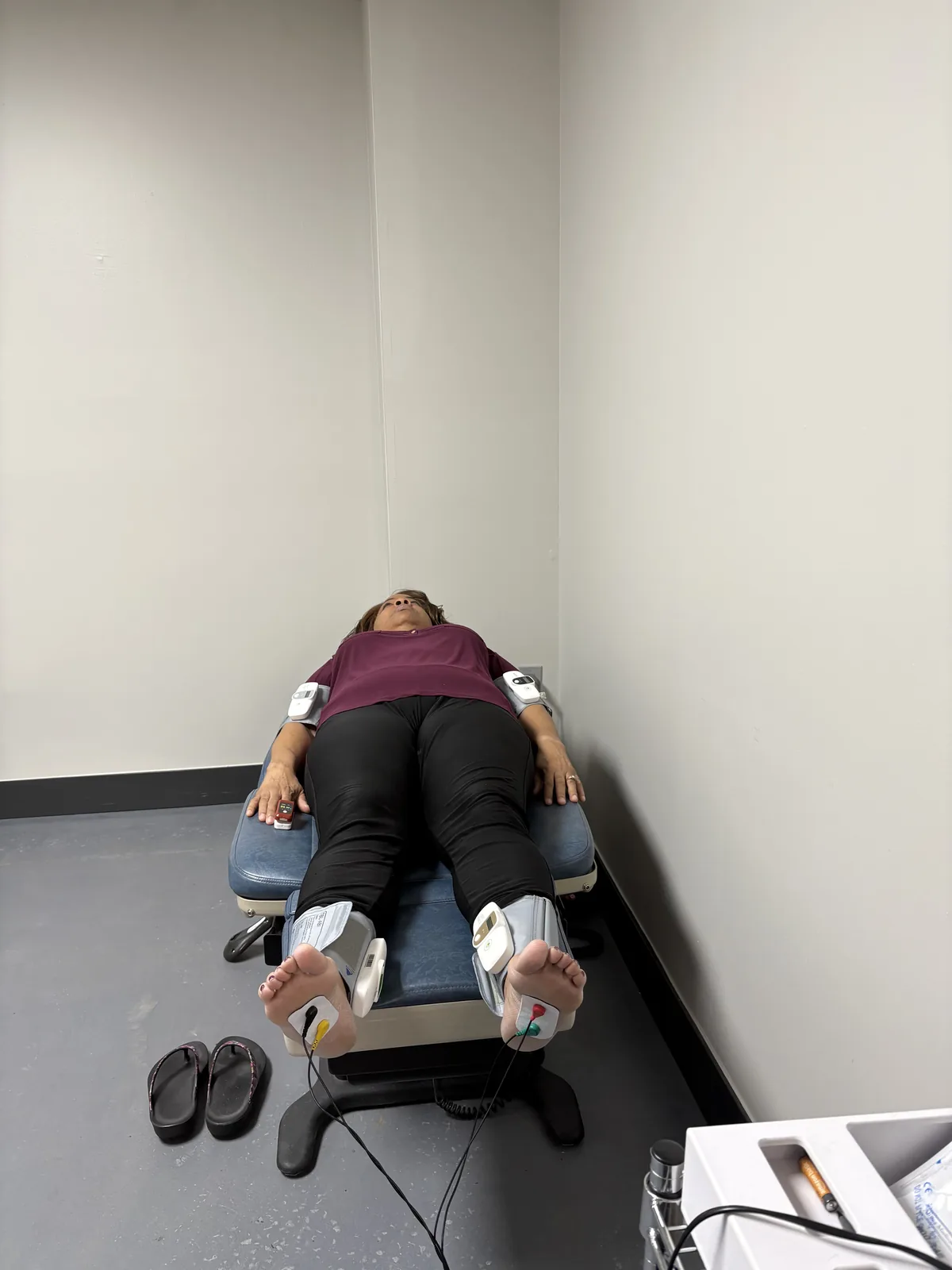
Nerve conduction studies and electromyography measure large myelinated fibers. By definition, those studies do not record small fibers. A patient with pure small fiber neuropathy can have severe burning every night and a fully normal electrodiagnostic study.1
This is worth being exact about, because patients are often told a normal result as reassurance. It is not reassurance. It is a negative finding on a test aimed somewhere else. The correct reading is that large fibers are intact. That is useful. It makes small-fiber-predominant disease more likely, not less.
Electrodiagnostic testing still belongs in the workup. It rules out large-fiber involvement. It finds any nerve compression that is also present. It sets a baseline. At Regenerve we do it on site. We always read it alongside the history, the exam and the search for a cause.
What test confirms small fiber neuropathy?
The reference standard is a 3 mm punch skin biopsy from the lower leg. The lab counts intraepidermal nerve fiber density (nerve fibers in the top layer of skin) and compares it with normal values for your age and sex. The European Federation of Neurological Societies and Peripheral Nerve Society guideline rates this as a reliable and efficient method of confirming the diagnosis.2 It is a small procedure done with local numbing. It gives a number that can be repeated later to measure change.
Two other tests show up in the workup. Quantitative sensory testing measures temperature and pain thresholds. Quantitative sudomotor axon reflex testing measures sweat responses driven by the small autonomic fibers.1 Both add information. Neither replaces the biopsy.
The cause is metabolic more often than “idiopathic”
A large share of small fiber neuropathy has long been labeled idiopathic (no known cause). Much of that label reflects incomplete testing.
In one study, 107 patients in a row with idiopathic neuropathy got a two-hour oral glucose tolerance test. Of them, 36 (34%) had impaired glucose tolerance. That is nearly three times the rate in controls of the same age. Fasting glucose and HbA1c were often normal in those patients. The OGTT was the test that found it. The authors concluded that impaired glucose tolerance may cause or contribute to small-fiber neuropathy with a phenotype resembling the painful sensory neuropathy seen in diabetes.3
The story does not end at glucose. One series looked at 219 patients with idiopathic neuropathy. Those with fully normal blood sugar carried the same high burden of metabolic syndrome features as those with impaired glucose tolerance. They also had significantly higher total and LDL cholesterol. Their HDL and triglycerides were more abnormal than in a comparison group with diabetes but no neuropathy.4
In other words, small fiber neuropathy tracks the whole metabolic terrain. That means glucose handling, lipids, blood pressure, central obesity (fat around the middle) and the supply from the tiniest blood vessels. It does not track any single lab value. Beyond the metabolic group, the list of possible causes includes autoimmune disease, B12 and other nutritional deficiencies, gluten-related nerve injury, thyroid disease, infections, toxic exposures, certain medications, and genetic sodium channel variants.1 The full list is in the 11 hidden drivers of peripheral neuropathy.
Small fiber density can change
This is the part worth holding onto. Small fiber density is measured in the skin, so it can be measured again. And it moves.
Thirty-two patients with impaired glucose tolerance and neuropathy got diet and exercise counseling built for them, for one year. Intraepidermal nerve fiber density rose by 1.4 fibers per millimeter at the proximal (upper) biopsy site. The gain correlated with less neuropathic pain.5 In a separate study, patients with diabetic peripheral neuropathy did a 10-week supervised aerobic and resistance exercise program. It produced significant reductions in pain and neuropathic symptoms. Biopsy also showed more intraepidermal nerve fiber branching.6
Both were small studies, and neither is a promise. But they show something that matters in the clinic. Treating the metabolic environment produces measurable change in the nerve fibers themselves. It is not just a change in how the patient rates their pain. That is the reasoning behind our approach, set out in the Regenerve Protocol. The training side is covered in exercises for peripheral neuropathy in the feet. The supplement evidence is graded in alternative medicine for peripheral neuropathy. That includes alpha-lipoic acid, which has been studied in exactly this symptom set.
What a proper evaluation should include
- A history and exam aimed at fiber type, not just at “does it hurt”.
- Electrodiagnostic testing to define large-fiber status and rule out compression.
- A two-hour oral glucose tolerance test, not a fasting glucose or HbA1c alone.
- A full lipid panel, read as part of the neuropathy workup and not as a separate heart issue.
- B12 with methylmalonic acid, thyroid studies, and screening for autoimmune and inflammatory causes.
- Skin biopsy for intraepidermal nerve fiber density when the diagnosis needs to be confirmed or measured.
- A medication review, including statins and long-term metformin.
Being evaluated in St. Louis
Regenerve is at 4477 Woodson Rd #104, St. Louis, MO 63134, minutes from St. Louis Lambert International Airport. It is led by Dr. Gurpreet Singh Padda, MD, MBA, MHP. The practice serves the St. Louis region, Missouri and Illinois.
Call (314) 886-5739 or text (314) 886-5902, or email info@regenerve.com. Office hours are Monday through Thursday 9:00 to 16:00 and Friday 9:00 to 13:30. Details are on the contact page and the locations page.
Did a nerve test come back normal while your feet still burn? The Nerve Damage Score is a five-question assessment. It produces a large-print report you can bring to any physician.
Burning that peaks after dark has its own list of causes. See burning feet at night: causes and evaluation.
Frequently asked questions
What are the symptoms of small fiber peripheral neuropathy?
Burning feet that are worse at night. Prickling and stabbing pain. Pain from light touch, such as bedding. Changed temperature feeling. Autonomic symptoms, including less sweating in the feet, lightheadedness on standing and gut changes. See peripheral neuropathy of the feet symptoms.
Is “small fibre neuropathy” the same as small fiber neuropathy?
Yes. “Small fibre” is the British spelling of the same condition. The diagnosis, testing and treatment are identical. See the hidden drivers of peripheral neuropathy.
Can small fiber neuropathy show up with a normal EMG and nerve conduction study?
Yes, and it often does. Those studies measure large myelinated fibers. So a pure small fiber neuropathy can cause severe symptoms with a normal result. See which specialist you should see.
How is small fiber neuropathy diagnosed?
By a 3 mm punch skin biopsy from the lower leg. The lab counts intraepidermal nerve fiber density against normal values. Quantitative sensory and sudomotor (sweat) testing back it up. See the Regenerve Protocol.
What causes small fiber neuropathy?
Metabolic causes lead. About a third of patients labeled idiopathic have impaired glucose tolerance that fasting glucose and HbA1c missed. Even patients with normal blood sugar carry a high burden of metabolic syndrome features, especially abnormal lipids. See how sugar damages nerves.
Can small nerve fibers grow back?
They can. Diet and exercise counseling over one year increased intraepidermal nerve fiber density on skin biopsy. The increase correlated with less neuropathic pain. Recovery is partial and slow, and no one can promise a cure. See exercises for peripheral neuropathy in the feet.
What are the treatment options for small fiber neuropathy?
Treatment starts with the cause, which is most often metabolic. In one study, a year of diet and exercise counseling increased small nerve fiber density on skin biopsy. Pain fell as the fibers returned. A separate 10-week supervised aerobic and resistance exercise program reduced pain and neuropathic symptoms. Supplements such as alpha-lipoic acid have also been studied for this symptom set. See the Regenerve Protocol.
Does small fiber neuropathy cause weakness?
Pure small fiber neuropathy usually does not. Strength, vibration sense and ankle reflexes usually stay normal. That is exactly what the diagnosis predicts, so normal strength does not rule it out. The main complaints are burning, prickling and pain from light touch. When weakness, lost vibration sense or absent reflexes appear, the larger nerve fibers have become involved too.
What blood tests look for the cause of small fiber neuropathy?
Start with a two-hour oral glucose tolerance test, because fasting glucose and HbA1c often read normal in people whose glucose handling is impaired. A full lipid panel belongs in the workup too. So do B12 with methylmalonic acid, thyroid studies, and screening for autoimmune and inflammatory causes. A review of medications, including statins and long-term metformin, rounds it out.
Sources
- Terkelsen AJ, et al. The diagnostic challenge of small fibre neuropathy: clinical presentations, evaluations, and causes. The Lancet Neurology. 2017. PMID 29029847
- Lauria G, et al. European Federation of Neurological Societies/Peripheral Nerve Society Guideline on the use of skin biopsy in the diagnosis of small fibre neuropathy. European Journal of Neurology. 2010. PMID 20642627
- Singleton JR, Smith AG, Bromberg MB. Increased prevalence of impaired glucose tolerance in patients with painful sensory neuropathy. Diabetes Care. 2001. PMID 11473085
- Smith AG, Rose K, Singleton JR. Idiopathic neuropathy patients are at high risk for metabolic syndrome. Journal of the Neurological Sciences. 2008. PMID 18606421
- Smith AG, et al. Lifestyle intervention for pre-diabetic neuropathy. Diabetes Care. 2006. PMID 16732011
- Kluding PM, et al. The effect of exercise on neuropathic symptoms, nerve function, and cutaneous innervation in people with diabetic peripheral neuropathy. Journal of Diabetes and its Complications. 2012. PMID 22717465