Neuropathy Resource Library

  • Exercises for Peripheral Neuropathy in the Feet: A Program Built on the Evidence

    Exercises for Peripheral Neuropathy in the Feet: A Program Built on the Evidence

    Exercise is the only treatment for peripheral neuropathy in the feet that has been shown, in people rather than animals, to change the nerve fibers themselves — and it is usually mentioned last, on the way out the door.

    This article sets out what the trials actually tested, why it works, and a practical weekly program you can start with, including the foot-protection rules that make exercising on numb feet safe.

    What the evidence says

    A 2022 systematic review with meta-analysis pooled 41 randomized controlled trials of exercise in patients with neuropathy, 27 of them in diabetic peripheral neuropathy. The authors concluded that evidence-based recommendations can now be made for this population, and that the most beneficial approach is a combination of endurance and sensorimotor training. Effects favored exercise across static balance, the Berg Balance Scale, the Timed Up and Go test, peroneal and sural nerve conduction velocity, and HbA1c.1

    Note what is in that list. Not just “patients felt better” — nerve conduction velocity and HbA1c both moved. Exercise is acting on the nerve and on the metabolic environment that damaged it.

    Two smaller studies show the same thing at the level of the nerve fiber. A 10-week supervised aerobic and resistance program in 17 people with diagnosed diabetic peripheral neuropathy produced significant reductions in pain and in neuropathic symptoms, together with increased intraepidermal nerve fiber branching on skin biopsy.2 And a year of individualized diet and exercise counseling in 32 patients with impaired glucose tolerance and neuropathy improved intraepidermal nerve fiber density by 1.4 fibers per millimeter at the proximal biopsy site, with the improvement correlating with reduced neuropathic pain.3

    These were small studies without large control groups, and they are not a promise of recovery. They are, however, direct evidence that nerve fibers respond to a change in the metabolic environment.

    Why it works: the metabolic explanation

    The nerves to your toes are the longest cells in the body, fed by the smallest blood vessels. They fail first when fuel and oxygen delivery falters. Exercise attacks that problem from several directions at once: it improves insulin sensitivity and glucose handling, lowers triglycerides, raises HDL, improves microvascular function, and increases blood flow to the vasa nervorum that supply the nerve itself.

    That breadth is exactly what this condition needs. Tightening glucose control alone has a clear preventive effect in type 1 diabetes but a much weaker one in type 2, where the lipid, blood pressure and insulin-resistance components of the terrain carry substantial weight.4 Among patients with idiopathic neuropathy, even those with entirely normal blood sugar carried a heavy burden of metabolic syndrome features, particularly abnormal lipids.5 Exercise is one of the few interventions that moves all of those at once. The mechanism is covered further in how sugar damages nerves.

    Before you start

    • Get cleared. Talk to your physician first, particularly if you have cardiac disease, retinopathy, kidney disease, an open wound, or an unexplained hot, swollen foot — the last of these needs evaluation before any weight-bearing exercise.
    • Inspect both feet before and after every session, including between the toes and the soles, using a mirror if you cannot see them easily. When protective sensation is reduced, the foot stops reporting injury, which is why the visual check replaces the pain you would normally feel.6
    • Wear properly fitted athletic shoes and seamless socks. Check inside the shoe with your hand before putting it on.
    • Have support available. Do balance work next to a counter, a wall or a sturdy chair.
    • Stop for any new blister, hot spot, unusual swelling, chest symptoms or a fall.

    A weekly program

    The evidence points to endurance plus sensorimotor training, with resistance work supporting both. Build to this over four to six weeks rather than starting at the top.

    1. Endurance — 3 to 5 days per week

    Start with 10 minutes and add 2 to 3 minutes per week, working toward 30 minutes at a pace where you can talk but not sing. Walking is the default. If foot pain, ulcer risk or balance make walking unwise, a stationary bike, a recumbent bike, a rowing machine, an arm ergometer or water walking deliver the same metabolic benefit while offloading the foot.

    2. Sensorimotor and balance work — 3 to 5 days per week, 10 to 15 minutes

    This is the component the meta-analysis identified as most crucial, and it is the one people skip. Always work within reach of a support.

    • Feet-together stand — 30 seconds, progressing to eyes closed once it is easy with eyes open
    • Semi-tandem then tandem stand — one foot partly, then fully in front of the other, 20 to 30 seconds each side
    • Single-leg stand — start at 5 to 10 seconds per side, build toward 30
    • Heel-to-toe walking — 10 steps along a countertop
    • Weight shifts — forward, back and side to side, slowly, feeling the pressure move across the foot
    • Head turns while standing — a harder challenge that trains the vestibular contribution to balance
    • Foam or cushion standing — only once firm-ground work is reliably steady

    Progress by removing input rather than by adding time: eyes open to eyes closed, two hands on the counter to one finger to none, firm surface to compliant surface.

    Two patients standing on whole-body vibration platforms in the therapy area at Regenerve, 4477 Woodson Rd, St. Louis, MO 63134

    3. Resistance — 2 to 3 days per week, non-consecutive

    Two to three sets of 10 to 15 repetitions of a small number of compound movements: sit-to-stand from a chair, heel raises, seated or standing leg press or wall squat, hamstring curls, hip abduction, and rows or presses for the upper body. Elastic-band resistance training has been studied specifically in older adults with diabetic peripheral neuropathy for its effect on balance and fear of falling, and it requires no equipment beyond a band.7

    4. Foot and ankle mobility — daily, 5 minutes

    • Ankle pumps, circles, and alphabet tracing with the toes
    • Toe curls and spreads, and picking up a towel with the toes
    • Calf and Achilles stretches — 30 seconds each side, twice
    • Seated rolling of the sole over a ball, gently, and never on numb skin you have not inspected

    5. Interrupt the sitting

    Stand and move for two to three minutes every 30 minutes. This is not filler. Uninterrupted sitting worsens glucose handling independently of whether you exercised earlier that day, and glucose handling is the thing you are trying to change.

    What to expect, and when

    Balance and steadiness usually improve first, often within four to six weeks, because they depend partly on training the remaining sensory input and the muscles that use it. Pain and burning tend to shift over two to three months. Changes at the level of nerve fibers were measured at 10 weeks in one study and at 12 months in the other — this is a long-horizon intervention.

    Some people notice a temporary increase in sensation or discomfort in the first weeks. That usually settles. Sharp new pain, a hot or swollen foot, or any open area means stop and be seen.

    Exercise is not the whole plan

    Exercise is the highest-yield thing you can do yourself. It does not replace finding out what is damaging the nerve. If the driver is a B12 deficiency, gluten-related injury, a medication, a toxic exposure or nerve compression, that needs to be addressed directly. The full list is in the 11 hidden drivers of peripheral neuropathy, how exercise fits alongside medical treatment is in peripheral neuropathy treatments for the feet, and the supplements people usually ask about next are graded in alternative medicine for peripheral neuropathy.

    Being evaluated in St. Louis

    Regenerve is at 4477 Woodson Rd #104, St. Louis, MO 63134, minutes from St. Louis Lambert International Airport, and is led by Dr. Gurpreet Singh Padda, MD, MBA, MHP. The practice serves the St. Louis region, Missouri and Illinois. Balance and gait training, physical rehabilitation and nutritional counseling are part of what we offer, and electrodiagnostic testing is performed on site. The full list is on the services page.

    Call or text (314) 886-5902, or email info@regenerve.com. Office hours are Monday through Thursday 9:00 to 16:00 and Friday 9:00 to 13:30. Details are on the contact page and the locations page.

    If you are not sure how far your neuropathy has progressed, the Nerve Damage Score is a five-question assessment that produces a large-print report you can bring to any physician.

    Frequently asked questions

    What are the best exercises for peripheral neuropathy in the feet?

    A combination of endurance training and sensorimotor or balance training, supported by resistance work. That combination is what a meta-analysis of 41 randomized trials identified as most beneficial in diabetic peripheral neuropathy. See peripheral neuropathy treatments for the feet.

    Is walking safe if my feet are numb?

    Usually yes, with properly fitted shoes and a visual inspection of both feet before and after every session, because a numb foot no longer reports injury. Any open wound or a hot, swollen foot needs evaluation before weight-bearing exercise. See peripheral neuropathy of the feet symptoms.

    Can exercise regrow damaged nerves?

    There is evidence that it can contribute. A 10-week supervised exercise program increased intraepidermal nerve fiber branching on skin biopsy, and a year of diet and exercise counseling increased nerve fiber density. Recovery is partial and slow, and no one can promise a cure. See small fiber neuropathy symptoms and testing.

    How long until exercise helps my neuropathy?

    Balance and steadiness often improve within four to six weeks. Pain and burning typically shift over two to three months, and changes measured at the nerve fiber were seen at 10 weeks and at 12 months in the published studies. See the Regenerve Protocol.

    What if I cannot walk far or my balance is poor?

    Use a seated or supported alternative for the endurance component — a recumbent bike, an arm ergometer or water walking — and do balance work holding a counter. The metabolic benefit does not require walking. See the hidden drivers of peripheral neuropathy.
    Should I exercise if it makes my feet hurt more?

    A temporary increase in sensation in the first weeks is common and usually settles. Sharp new pain, a new blister or hot spot, or a hot and swollen foot means stop and be evaluated. See which specialist you should see.

    Sources

    1. Streckmann F, et al. Exercise and Neuropathy: Systematic Review with Meta-Analysis. Sports Medicine. 2022. PMID 34964950
    2. Kluding PM, et al. The effect of exercise on neuropathic symptoms, nerve function, and cutaneous innervation in people with diabetic peripheral neuropathy. Journal of Diabetes and its Complications. 2012. PMID 22717465
    3. Smith AG, et al. Lifestyle intervention for pre-diabetic neuropathy. Diabetes Care. 2006. PMID 16732011
    4. Callaghan BC, Little AA, Feldman EL, Hughes RA. Enhanced glucose control for preventing and treating diabetic neuropathy. Cochrane Database of Systematic Reviews. 2012. PMID 22696371
    5. Smith AG, Rose K, Singleton JR. Idiopathic neuropathy patients are at high risk for metabolic syndrome. Journal of the Neurological Sciences. 2008. PMID 18606421
    6. Boulton AJM, et al. Comprehensive foot examination and risk assessment: a report of the task force of the foot care interest group of the American Diabetes Association. Diabetes Care. 2008. PMID 18663232
    7. Armat MR, et al. The Effect of Resistance Exercises Using an Elastic Band on Balance and Fear of Falling in Older Adults With Diabetic Peripheral Neuropathy. Archives of Physical Medicine and Rehabilitation. 2024. PMID 38218307
  • Alternative Medicine for Peripheral Neuropathy: What the Evidence Actually Supports

    Alternative Medicine for Peripheral Neuropathy: What the Evidence Actually Supports

    People look for alternative treatments for peripheral neuropathy for a reasonable reason: the standard drugs treat the pain and not the nerve, and their average effect is modest. That is a real gap, and it is worth filling. It is also the gap that the supplement industry has filled with a great deal of confident marketing and very little published data.

    What follows is a grading of the non-drug options by the evidence that actually exists — what has randomized trial support, what has weak support, what has failed when properly tested, and the one intervention that outperforms every bottle on the shelf.

    Start with what the label “alternative” is hiding

    Most people searching for alternative medicine for neuropathy have already been told there is nothing to be done about the cause. In a large share of cases that is not true — the cause simply was not looked for thoroughly enough.

    Among 107 patients whose neuropathy had been called idiopathic, 34% had impaired glucose tolerance, close to three times the rate in matched controls, and in many of them fasting glucose and HbA1c were normal. Only a two-hour oral glucose tolerance test found it.1 In a separate series of 219 idiopathic neuropathy patients, even those with entirely normal blood sugar carried a heavy burden of metabolic syndrome features, with lipid abnormalities particularly prevalent.2

    So before spending money on supplements, it is worth confirming that the workup included an OGTT, a full lipid panel, B12 with methylmalonic acid, thyroid studies, and a review of medications. The 11 hidden drivers of peripheral neuropathy covers the rest of that list, and if your nerve conduction study came back normal while your feet still burn, read small fiber peripheral neuropathy symptoms and testing before concluding that nothing was found.

    Cardiometabolic vascular elasticity report showing vascular, endothelial, autonomic and sweat-response assessment used in the neuropathy workup at Regenerve, 4477 Woodson Rd, St. Louis, MO 63134

    The strongest non-drug intervention is not a supplement

    Diet and exercise directed at the metabolic terrain has better evidence than anything sold as a nerve supplement — and it is measured at the nerve, not on a questionnaire.

    Thirty-two patients with impaired glucose tolerance and neuropathy received individualized diet and exercise counseling for one year, with skin biopsies at baseline and at 12 months. Intraepidermal nerve fiber density improved by 1.4 fibers per millimeter at the proximal site, and that improvement correlated with reduced neuropathic pain.3 A separate 10-week supervised aerobic and resistance program in patients with diagnosed diabetic peripheral neuropathy produced significant reductions in pain and neuropathic symptoms alongside increased intraepidermal nerve fiber branching on biopsy.4

    A 2022 systematic review with meta-analysis of 41 randomized controlled trials concluded that for diabetic peripheral neuropathy the evidence now supports a specific recommendation: a combination of endurance and sensorimotor training, with benefits across balance measures, functional mobility, peroneal and sural nerve conduction velocity, and HbA1c.5

    Nothing in the supplement aisle has that level of support. The practical program is in exercises for peripheral neuropathy in the feet.

    Alpha-lipoic acid: real evidence, with an important qualifier

    Alpha-lipoic acid is an antioxidant and the best-studied supplement in this field. A meta-analysis of four randomized, double-blind, placebo-controlled trials — ALADIN I, ALADIN III, SYDNEY and NATHAN II, totaling 1,258 patients — found a 24.1% relative improvement in the Total Symptom Score in the feet versus placebo after three weeks, with responder rates of 52.7% on alpha-lipoic acid against 36.9% on placebo. Pain, burning and numbness all improved, as did pinprick and touch-pressure sensation and ankle reflexes, with no difference in adverse event rates.6

    The qualifier matters: that evidence is for 600 mg given intravenously, daily for three weeks. The oral capsules sold over the counter are not the same intervention, and their evidence is considerably weaker. If a supplement is recommended to you on the strength of “the ALADIN trials,” it is fair to ask whether the study used the oral form.

    Acetyl-L-carnitine: modest, in a specific population

    Acetyl-L-carnitine improved pain, vibratory perception and measures of nerve regeneration in patients with chronic diabetic neuropathy in randomized trial data, with the clearest effect in those treated earlier in the disease course.7 It is a reasonable adjunct with a real, if modest, evidence base. It is not a substitute for treating the driver.

    Palmitoylethanolamide: promising, and worth reading carefully

    Palmitoylethanolamide, or PEA, is a naturally occurring fatty acid amide. A systematic review and meta-analysis of 11 double-blind randomized controlled trials covering 774 patients with chronic pain found reduced pain scores relative to comparators, with no major side effects attributed to PEA in any study.8 The authors noted that optimal dosing and administration still need to be determined, and the trials pooled were across chronic pain generally rather than neuropathy alone.

    PEA is one of the supportive agents we use at our St. Louis clinic, within a plan rather than as a standalone answer.

    Acupuncture: low-certainty evidence, genuine effect size

    A 2025 systematic review and meta-analysis of 14 randomized trials covering 1,169 participants found that, compared with sham, acupuncture may reduce pain in diabetic peripheral neuropathy by 1.44 cm on a 10 cm visual analog scale, and may reduce overall neurologic symptom severity, with little to no difference in adverse events. The authors graded the certainty of that evidence as low.9
    Low certainty does not mean it does not work. It means the trials were small or at risk of bias and the estimate could move. Given the safety profile, acupuncture is a defensible thing to try alongside cause-directed treatment.

    Topical capsaicin: the one that may do more than mask

    High-concentration capsaicin at 8% is a prescription patch rather than a store-bought cream, but people often find it while searching for natural options because capsaicin comes from chili peppers. It works by defunctionalizing overactive nociceptive fibers in the skin, and there is published evidence of subsequent nerve fiber regeneration accompanying the pain relief.10 We use it, and it is covered in the high-concentration capsaicin protocol.

    Light-based therapies: mechanistic rationale, thin trial evidence

    Photobiomodulation targets cytochrome c oxidase in mitochondria and is intended to support energy production and circulation in injured nerve tissue. We offer class 4 photobiomodulation, class 3B cold laser and whole-body infrared at the clinic, and describe them on the services page.

    The honest statement of the evidence is that the mechanistic rationale is coherent and the randomized trial base for diabetic peripheral neuropathy is still limited — several systematic reviews have been registered and published as protocols rather than completed syntheses. We use these therapies as adjuncts within a plan built on identifying and treating the cause, and we do not present them as the treatment.

    What has failed when properly tested

    This section matters as much as the others. Benfotiamine, a fat-soluble thiamine derivative sold widely for neuropathy, was tested in a 12-month randomized, placebo-controlled, double-blind trial in type 2 diabetes patients with symptomatic polyneuropathy. It raised blood levels of all six thiamine analytes measured, confirming it was absorbed and doing something biochemically. It produced no significant effect on corneal nerve fiber length, on the secondary morphometric, functional and clinical neuropathy outcomes, or on quality of life.11

    That is what a well-conducted negative trial looks like, and it is a useful benchmark. Ask of any product marketed for neuropathy: has it been tested this way, and what happened?

    Things worth being skeptical about

    • Any product or program promising to cure, reverse or eliminate neuropathy. Nerve recovery is partial, slow and dependent on the cause. A cure claim is a marketing claim.
    • Proprietary “nerve support” blends that combine a dozen ingredients at doses below those used in any trial, so no individual component reaches a studied dose.
    • Fixed packages of sessions sold before a diagnosis. The number of treatments should follow clinical need and response, not a package purchased in advance.
    • High-dose vitamin B6. Excess B6 causes a sensory neuropathy in its own right. More is not better here.
    • Anything that requires stopping your prescribed medication to work.

    How we think about this

    Alternative and complementary approaches are not a separate category of medicine to us. They are treatments with an evidence base to be graded like any other. Where the evidence supports something, we use it; where it does not, we say so; and none of it substitutes for identifying what is damaging the nerve. That sequencing is set out in the Regenerve Protocol, and the full treatment landscape is in peripheral neuropathy treatments for the feet.

    Talk to your physician before starting any supplement, particularly if you take anticoagulants, thyroid medication or diabetes medication, and note that alpha-lipoic acid can lower blood glucose.

    Being evaluated in St. Louis

    Regenerve is at 4477 Woodson Rd #104, St. Louis, MO 63134, minutes from St. Louis Lambert International Airport, and is led by Dr. Gurpreet Singh Padda, MD, MBA, MHP. The practice serves the St. Louis region, Missouri and Illinois. Electrodiagnostic testing is performed on site.

    Call or text (314) 886-5902, or email info@regenerve.com. Office hours are Monday through Thursday 9:00 to 16:00 and Friday 9:00 to 13:30. Details are on the contact page and the locations page.

    Before buying anything, the Nerve Damage Score is a five-question assessment that produces a large-print report you can bring to any physician.

    Frequently asked questions

    What is the best alternative treatment for peripheral neuropathy?

    Diet and exercise aimed at the metabolic terrain has the strongest evidence, including measured regrowth of nerve fibers on skin biopsy. No supplement matches it. See exercises for peripheral neuropathy in the feet.

    Does alpha-lipoic acid work for neuropathy?

    The strong trial evidence is for 600 mg given intravenously over three weeks, which produced a 24.1% relative improvement in symptom scores versus placebo. Evidence for over-the-counter oral capsules is considerably weaker. See how sugar damages nerves.

    Does acupuncture help neuropathy in the feet?

    A meta-analysis of 14 randomized trials found acupuncture may reduce pain and neurologic symptom severity compared with sham, with low certainty of evidence and few adverse events. See peripheral neuropathy treatments for the feet.

    Should I take benfotiamine for neuropathy?

    A 12-month randomized placebo-controlled trial found benfotiamine raised thiamine levels but had no significant effect on nerve structure, nerve function, clinical neuropathy measures or quality of life. See functional B12 deficiency and neuropathy.

    Are supplements enough on their own?

    No. Supplements address one input at a time, while neuropathy is driven by the whole metabolic environment plus any nutritional, autoimmune, toxic or structural driver present. See the hidden drivers of peripheral neuropathy.

    Can vitamins make neuropathy worse?

    Yes. High-dose vitamin B6 can cause a sensory neuropathy of its own, so supplementing without knowing your levels can add a driver rather than remove one. See functional B12 deficiency and neuropathy.

    Sources

    1. Singleton JR, Smith AG, Bromberg MB. Increased prevalence of impaired glucose tolerance in patients with painful sensory neuropathy. Diabetes Care. 2001. PMID 11473085
    2. Smith AG, Rose K, Singleton JR. Idiopathic neuropathy patients are at high risk for metabolic syndrome. Journal of the Neurological Sciences. 2008. PMID 18606421
    3. Smith AG, et al. Lifestyle intervention for pre-diabetic neuropathy. Diabetes Care. 2006. PMID 16732011
    4. Kluding PM, et al. The effect of exercise on neuropathic symptoms, nerve function, and cutaneous innervation in people with diabetic peripheral neuropathy. Journal of Diabetes and its Complications. 2012. PMID 22717465
    5. Streckmann F, et al. Exercise and Neuropathy: Systematic Review with Meta-Analysis. Sports Medicine. 2022. PMID 34964950
    6. Ziegler D, Nowak H, Kempler P, Vargha P, Low PA. Treatment of symptomatic diabetic polyneuropathy with the antioxidant alpha-lipoic acid: a meta-analysis. Diabetic Medicine. 2004. PMID 14984445
    7. Sima AAF, et al. Acetyl-L-carnitine improves pain, nerve regeneration, and vibratory perception in patients with chronic diabetic neuropathy. Diabetes Care. 2005. PMID 15616239
    8. Lang-Illievich K, et al. Palmitoylethanolamide in the Treatment of Chronic Pain: A Systematic Review and Meta-Analysis of Double-Blind Randomized Controlled Trials. Nutrients. 2023. PMID 36986081
    9. Lan L, et al. Acupuncture for the Management of Chronic Diabetic Peripheral Neuropathy: A Systematic Review and Meta-Analysis of Randomized Controlled Trials. Current Pain and Headache Reports. 2025. PMID 40220243
    10. Anand P, et al. Reversing painful and non-painful diabetic neuropathy with the capsaicin 8% patch: clinical evidence for pain relief and restoration of function via nerve fiber regeneration. Frontiers in Neurology. 2022. PMID 36388188
    11. Ziegler D, et al. Effects of benfotiamine treatment over 12 months on morphometric, neurophysiological and clinical measures in type 2 diabetes patients with symptomatic polyneuropathy (BOND study). BMJ Open Diabetes Research & Care. 2026. PMID 41571333
  • Small Fiber Peripheral Neuropathy: Symptoms, Testing, and the Metabolic Cause Behind Most Cases

    Small Fiber Peripheral Neuropathy: Symptoms, Testing, and the Metabolic Cause Behind Most Cases

    Small fiber neuropathy is the diagnosis most often missed in people whose feet burn. The symptoms are severe, the standard nerve test is frequently normal, and the patient is sent home with the impression that nothing was found. Nothing was found because the test that was run does not look at the fibers that are damaged.

    This article covers what the symptoms actually are, how the condition is properly tested for, and why the cause — in a large share of cases — turns out to be metabolic.

    What “small fiber” means

    Peripheral sensory nerves come in two calibers. Large myelinated fibers carry vibration, light touch, position sense and the signals behind your ankle reflexes. Small fibers — thinly myelinated A-delta and unmyelinated C fibers — carry pain, temperature, and the autonomic traffic that controls sweating, blood vessel tone and internal organ function.1

    Small fibers are the most metabolically exposed part of the system. They are unmyelinated or barely myelinated, they terminate in the skin far from their cell bodies, and they depend on the smallest blood vessels in the body. When the metabolic environment deteriorates, they fail first — and they fail at the toes first, because that is where the longest ones end.

    The symptoms

    Sensory symptoms

    • Burning feet, the most characteristic complaint, typically worse at night and worse at rest
    • Pins and needles, prickling or tingling that does not resolve with movement
    • Sharp, stabbing or electric pain in bursts
    • Allodynia — the weight of bedding on the toes registering as pain
    • Altered temperature sensation, where cold feels burning or hot water cannot be judged reliably
    • A sense of walking on pebbles, sand, or bunched socks that are not there

    The distribution is symmetrical and length-dependent: toes first, then feet, then ascending the legs over months to years, with the hands involved only much later.

    Autonomic symptoms

    This is the half that rarely gets connected to the feet, because the symptoms appear elsewhere in the body and get reported to different specialists:

    • Reduced or absent sweating in the feet, leaving skin dry, cracked and shiny — or, less often, excessive sweating elsewhere
    • Lightheadedness on standing from impaired blood vessel constriction
    • Color and temperature changes in the feet — mottling, redness, or feet that feel cold to you and warm to the touch
    • Gastrointestinal changes: early fullness after eating, bloating, constipation or diarrhea
    • Bladder changes and erectile dysfunction
    • Dry eyes and dry mouth

    When burning feet and several of these appear together, small fiber neuropathy is a far better explanation than five unrelated diagnoses.1

    What is usually absent

    Pure small fiber neuropathy generally spares strength, vibration sense and reflexes. Normal ankle reflexes and normal strength do not argue against the diagnosis — they are what the diagnosis predicts. Once weakness, lost vibration sense and absent reflexes appear, large fibers have been recruited into the process as well.

    Why standard nerve testing comes back normal

    Nerve conduction studies and electromyography measure large myelinated fibers. Small fibers are, by definition, not what those studies record. A patient with pure small fiber neuropathy can have severe nightly burning and an entirely normal electrodiagnostic study.1

    This is worth being precise about, because a normal result is often reported to the patient as reassurance. It is not reassurance. It is a negative finding on a test aimed elsewhere. The correct interpretation is that large fibers are intact, which is useful information — it makes small-fiber-predominant disease more likely, not less.

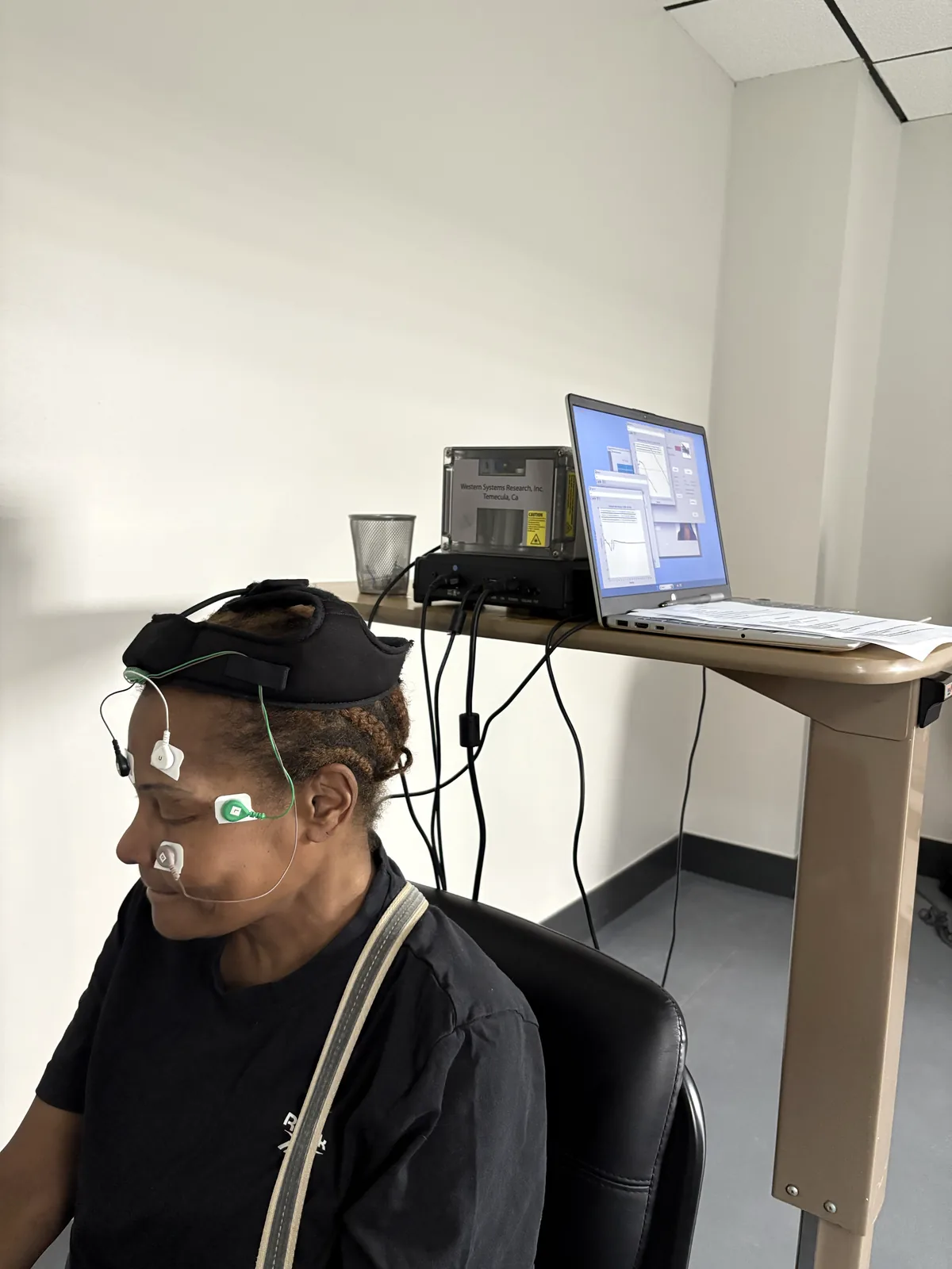
    Electrodiagnostic testing still belongs in the workup. It rules out large-fiber involvement, identifies coexisting nerve compression, and establishes a baseline. At Regenerve it is performed on site, and it is always interpreted alongside the history, the examination and the search for a cause.

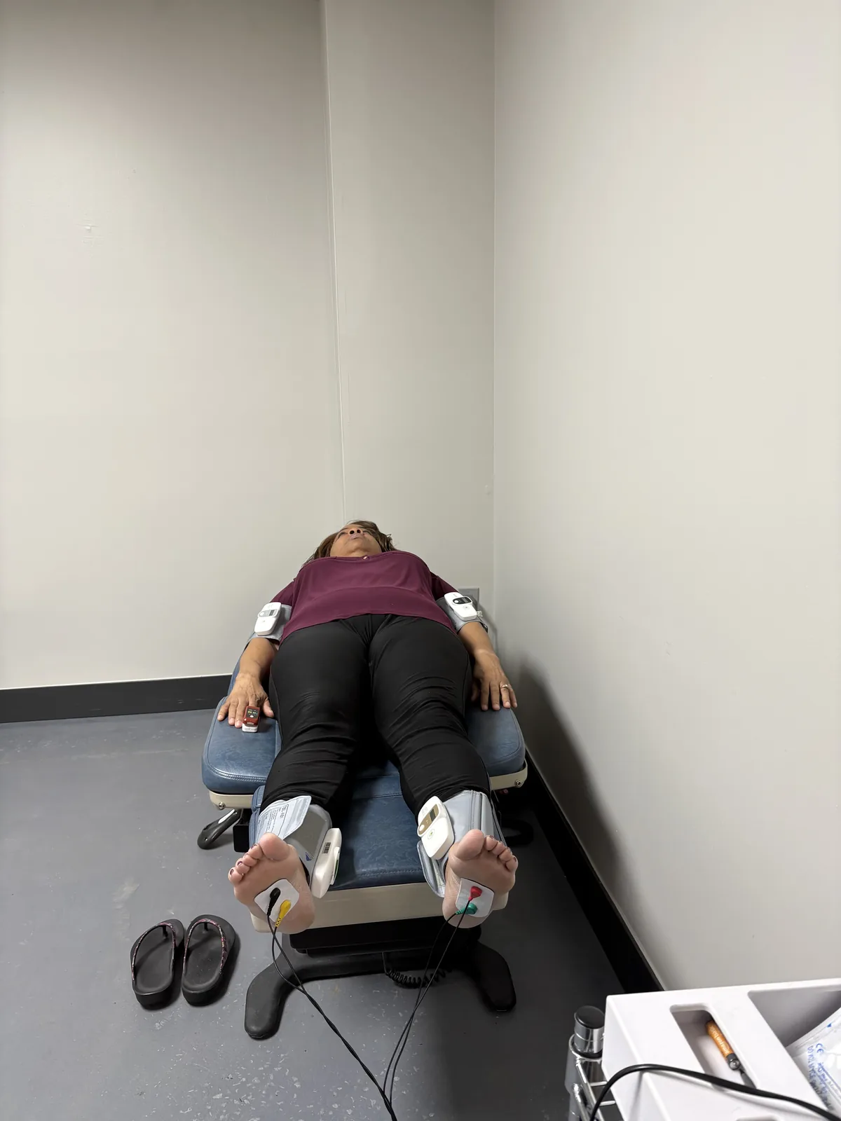
    Patient reclined for small-fiber and autonomic nerve testing, with sensor cuffs on both ankles and wrists, at Regenerve, 4477 Woodson Rd, St. Louis, MO 63134

    How small fiber neuropathy is confirmed

    The reference standard is a 3 mm punch skin biopsy from the distal leg, with intraepidermal nerve fiber density counted against age- and sex-matched normative values. The European Federation of Neurological Societies and Peripheral Nerve Society guideline rates this as a reliable and efficient method of confirming the diagnosis.2 It is a small procedure done under local anesthetic, and it produces a number that can be repeated later to measure change.

    Two other tests appear in the workup. Quantitative sensory testing measures thermal and pain thresholds, and quantitative sudomotor axon reflex testing measures sweat responses driven by the small autonomic fibers.1 Both add information; neither replaces the biopsy.

    The cause is metabolic more often than “idiopathic”

    A large proportion of small fiber neuropathy has historically been labeled idiopathic. Much of that label reflects incomplete testing.

    When 107 consecutive patients with idiopathic neuropathy were given a two-hour oral glucose tolerance test, 36 of them — 34% — had impaired glucose tolerance, nearly three times the rate in age-matched controls. Fasting glucose and HbA1c were frequently normal in those patients; the OGTT was the test that found it. The authors concluded that impaired glucose tolerance may cause or contribute to small-fiber neuropathy with a phenotype resembling the painful sensory neuropathy seen in diabetes.3

    The story does not end at glucose. In a series of 219 patients with idiopathic neuropathy, those with completely normal blood sugar carried the same elevated burden of metabolic syndrome features as those with impaired glucose tolerance, and had significantly higher total and LDL cholesterol and more abnormal HDL and triglycerides than a comparison group with diabetes but no neuropathy.4

    In other words: small fiber neuropathy tracks the metabolic terrain as a whole — glucose handling, lipids, blood pressure, central obesity and the microvascular supply — not any single laboratory value. Beyond the metabolic group, the differential includes autoimmune disease, B12 and other nutritional deficiencies, gluten-related nerve injury, thyroid disease, infections, toxic exposures, certain medications, and genetic sodium channel variants.1 The full list is in the 11 hidden drivers of peripheral neuropathy.

    Small fibers can regrow

    This is the part worth holding onto. Because small fiber density is measured in the skin, it can be measured again — and it moves.

    Thirty-two patients with impaired glucose tolerance and neuropathy received individualized diet and exercise counseling for one year. Intraepidermal nerve fiber density improved by 1.4 fibers per millimeter at the proximal biopsy site, and the improvement correlated with reduced neuropathic pain.5 Separately, a 10-week supervised aerobic and resistance exercise program in patients with diabetic peripheral neuropathy produced significant reductions in pain and neuropathic symptoms alongside increased intraepidermal nerve fiber branching on biopsy.6

    Both were small studies, and neither is a promise. But they demonstrate something that matters clinically: treating the metabolic environment produces measurable change in the nerve fibers themselves, not merely in how the patient rates their pain. That is the reasoning behind our approach, set out in the Regenerve Protocol. The training side is covered in exercises for peripheral neuropathy in the feet, and the supplement evidence — including alpha-lipoic acid, which has been studied in exactly this symptom set — is graded in alternative medicine for peripheral neuropathy.

    What a proper evaluation should include

    • A history and examination oriented to fiber type, not just to “does it hurt”
    • Electrodiagnostic testing to define large-fiber status and exclude compression
    • A two-hour oral glucose tolerance test, not a fasting glucose or HbA1c alone
    • A full lipid panel, read as part of the neuropathy workup rather than a separate cardiac issue
    • B12 with methylmalonic acid, thyroid studies, and screening for autoimmune and inflammatory causes
    • Skin biopsy for intraepidermal nerve fiber density where the diagnosis needs confirming or quantifying
    • A medication review, including statins and long-term metformin

    Being evaluated in St. Louis

    Regenerve is at 4477 Woodson Rd #104, St. Louis, MO 63134, minutes from St. Louis Lambert International Airport, and is led by Dr. Gurpreet Singh Padda, MD, MBA, MHP. The practice serves the St. Louis region, Missouri and Illinois.

    Call or text (314) 886-5902, or email info@regenerve.com. Office hours are Monday through Thursday 9:00 to 16:00 and Friday 9:00 to 13:30. Details are on the contact page and the locations page.

    If a nerve test came back normal and your feet still burn, the Nerve Damage Score is a five-question assessment that produces a large-print report you can bring to any physician.

    Frequently asked questions

    What are the symptoms of small fiber peripheral neuropathy?

    Burning feet that are worse at night, prickling and stabbing pain, pain from light touch such as bedding, altered temperature sensation, and autonomic symptoms including reduced sweating in the feet, lightheadedness on standing and gastrointestinal changes. See peripheral neuropathy of the feet symptoms.

    Is “small fibre neuropathy” the same as small fiber neuropathy?

    Yes. “Small fibre” is the British spelling of the same condition; the diagnosis, testing and treatment are identical. See the hidden drivers of peripheral neuropathy.

    Can small fiber neuropathy show up with a normal EMG and nerve conduction study?

    Yes, and it commonly does. Those studies measure large myelinated fibers, so a pure small fiber neuropathy can produce severe symptoms with a normal result. See which specialist you should see.

    How is small fiber neuropathy diagnosed?

    By a 3 mm punch skin biopsy from the distal leg with intraepidermal nerve fiber density counted against normative values, supported by quantitative sensory and sudomotor testing. See the Regenerve Protocol.

    What causes small fiber neuropathy?

    Metabolic causes lead. About a third of patients labeled idiopathic have impaired glucose tolerance that fasting glucose and HbA1c missed, and even normoglycemic patients carry a high burden of metabolic syndrome features, particularly abnormal lipids. See how sugar damages nerves.

    Can small nerve fibers grow back?

    They can. Diet and exercise counseling over one year increased intraepidermal nerve fiber density on skin biopsy, and the increase correlated with less neuropathic pain. Recovery is partial and slow, and no one can promise a cure. See exercises for peripheral neuropathy in the feet.

    Sources

    1. Terkelsen AJ, et al. The diagnostic challenge of small fibre neuropathy: clinical presentations, evaluations, and causes. The Lancet Neurology. 2017. PMID 29029847
    2. Lauria G, et al. European Federation of Neurological Societies/Peripheral Nerve Society Guideline on the use of skin biopsy in the diagnosis of small fibre neuropathy. European Journal of Neurology. 2010. PMID 20642627
    3. Singleton JR, Smith AG, Bromberg MB. Increased prevalence of impaired glucose tolerance in patients with painful sensory neuropathy. Diabetes Care. 2001. PMID 11473085
    4. Smith AG, Rose K, Singleton JR. Idiopathic neuropathy patients are at high risk for metabolic syndrome. Journal of the Neurological Sciences. 2008. PMID 18606421
    5. Smith AG, et al. Lifestyle intervention for pre-diabetic neuropathy. Diabetes Care. 2006. PMID 16732011
    6. Kluding PM, et al. The effect of exercise on neuropathic symptoms, nerve function, and cutaneous innervation in people with diabetic peripheral neuropathy. Journal of Diabetes and its Complications. 2012. PMID 22717465
  • Peripheral Neuropathy of the Feet: Symptoms, Patterns, and What They Mean

    Peripheral Neuropathy of the Feet: Symptoms, Patterns, and What They Mean

    Peripheral neuropathy in the feet does not announce itself. It arrives as something easy to explain away — socks that feel bunched when they are not, a floor that feels slightly wrong underfoot, toes that burn at night and are fine by morning. By the time most people describe it to a physician, the process has usually been running for years.

    The symptoms are worth reading carefully, because their pattern tells you a great deal about what is damaging the nerve and how far it has gone.

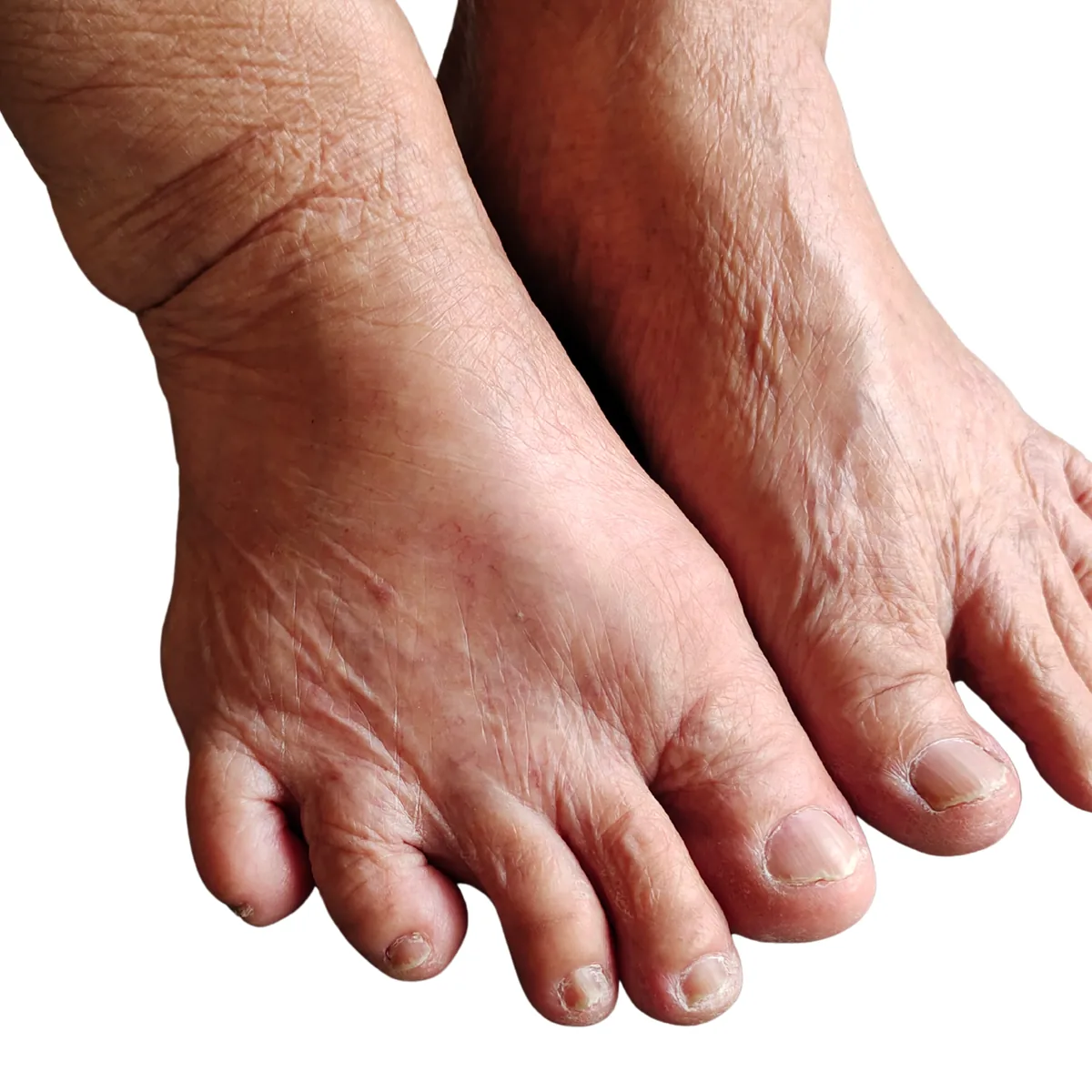
    Bare feet of an older adult showing dry skin, thickened toenails and bunion deformity, the appearance often seen alongside peripheral neuropathy of the feet

    The pattern matters more than any single symptom

    Metabolic neuropathy is length-dependent. It affects the longest nerve fibers first, which means it starts at the toes, spreads upward symmetrically on both sides, and only reaches the fingertips once it has climbed to roughly knee height. Clinicians call this a stocking-glove distribution, and it is the signature of a systemic process affecting every nerve at once rather than an injury to one nerve.1

    Two features of that pattern carry real diagnostic weight. It is symmetrical — both feet, roughly equally. And it ascends. Symptoms in one foot only, or symptoms that skip from a foot to a hand without covering the ground between, point somewhere else, often toward nerve compression or a double-crush pattern.

    Positive symptoms: what the nerve adds

    Damaged sensory nerves misfire, generating signals with no stimulus behind them. Patients describe these in remarkably consistent language:

    • Burning, often described as the feet being on fire, and often worst at night when there is nothing else competing for attention
    • Tingling or pins and needles that does not resolve when you move the foot
    • Electric or shooting pain arriving in bursts, sometimes without any trigger
    • Allodynia — pain from something that should not hurt, most classically the weight of a bedsheet on the toes
    • Deep aching in the arches or across the ball of the foot
    • Crawling, itching or wetness where nothing is touching the skin

    These are the symptoms that bring people in, and they are also the ones most likely to be treated in isolation. Relief matters. But a burning foot is a report about the nerve’s condition, and turning down the volume does not change what generated it.

    Negative symptoms: what the nerve stops delivering

    The quieter half of the picture is loss, and it is the more dangerous half:

    • Numbness, or a sense that the feet are wrapped in a layer of something
    • Reduced ability to feel temperature, which is why burns from hot water and heating pads are a genuine risk
    • Loss of position sense, so you no longer know where your feet are without looking at them
    • Unsteadiness, especially in the dark or on uneven ground — a very common first complaint
    • Weakness in the foot and ankle, sometimes showing up as tripping on curbs or scuffing a toe
    • Not noticing injury — the blister, the stone in the shoe, the crack in the heel

    That last item is the one that changes the stakes. When protective sensation is gone, the foot no longer reports damage, and a minor injury can progress without ever hurting. A 10-gram monofilament test identifies loss of protective sensation, and anyone who has it needs daily visual foot checks and well-fitted footwear as a standing routine.2

    Small fibers and large fibers give different symptoms

    Sensory nerves come in two calibers, and they fail differently. Small unmyelinated and thinly myelinated fibers carry pain, temperature and autonomic signals. Large myelinated fibers carry vibration, light touch and position sense.

    Small-fiber symptoms come first in most metabolic neuropathy: burning, pinprick pain, altered temperature sensation, and autonomic changes such as feet that no longer sweat, skin that has become dry and shiny, or color changes. Large-fiber symptoms arrive later: numbness, lost vibration sense, imbalance, and reduced or absent ankle reflexes.3

    This distinction has a practical consequence that catches a lot of patients. Standard nerve conduction studies measure large fibers. If your neuropathy is still predominantly small-fiber, those studies can come back normal while your feet are burning every night — and a normal result gets misread as “nothing is wrong.” It means the test looked at the wrong fibers. We cover this in detail in small fiber peripheral neuropathy symptoms and testing.

    Autonomic symptoms people rarely connect to their feet

    The same small fibers that carry pain also control sweating, blood vessel tone and internal organ function. When they are damaged, symptoms appear well beyond the feet: lightheadedness on standing, unexplained sweating changes, early fullness after meals, constipation or diarrhea, bladder changes, and erectile dysfunction. These are often reported to different specialists as separate problems. They frequently belong to one process.

    What the symptoms are usually telling you

    Peripheral neuropathy is not rare and it is not confined to people with diabetes. In a national sample of U.S. adults aged 40 and older, 13.5% had peripheral neuropathy — 27.0% of those with diabetes, and 11.6% of those without. In both groups it was independently associated with mortality over a median 13 years of follow-up.4 See diabetic peripheral neuropathy treatment in St. Louis.

    The metabolic connection runs deeper than a diabetes diagnosis. Among patients whose neuropathy had been labeled idiopathic, 34% turned out to have impaired glucose tolerance — about three times the rate in age-matched controls — and in many of them the fasting glucose and HbA1c were normal, with only a two-hour glucose tolerance test revealing the problem.5 A separate series found that even the normoglycemic patients with idiopathic neuropathy carried a heavy load of metabolic syndrome features, with lipid abnormalities especially prevalent.6

    So when the feet start burning, the useful question is not only “how bad is the nerve damage” but “what is the metabolic environment that produced it.” Those symptoms are frequently the first visible output of a systemic problem that has not yet been named. The mechanism is covered in how sugar damages nerves.

    When symptoms need attention sooner rather than later

    Most feet-first neuropathy develops over years. Some presentations should not wait:

    • Rapid progression over days or weeks
    • Marked weakness rather than mainly sensory symptoms
    • Symptoms that are clearly asymmetrical, or confined to one nerve’s territory
    • Symptoms beginning in the hands and feet at the same time
    • Any open wound, ulcer, unexplained swelling, redness or warmth in a numb foot
    • New bladder or bowel changes alongside the foot symptoms

    The last of these can indicate something above the peripheral nerve entirely and warrants prompt evaluation.

    How the symptoms get turned into a diagnosis

    “You have neuropathy” is a restatement of your symptoms, not an answer. A useful evaluation establishes three things: which fibers are affected, how far the damage has progressed, and what is driving it. That means a proper neurologic examination, electrodiagnostic testing where large-fiber involvement is in question, and a genuine search for the driver — metabolic, nutritional, autoimmune, toxic, pharmacologic or structural. At Regenerve, electrodiagnostic testing is performed on site. The full range of drivers worth ruling out is in the 11 hidden drivers of peripheral neuropathy, what treatment follows is in peripheral neuropathy treatments for the feet, and the non-drug options are graded in alternative medicine for peripheral neuropathy.

    Being evaluated in St. Louis

    Regenerve is at 4477 Woodson Rd #104, St. Louis, MO 63134, minutes from St. Louis Lambert International Airport, and is led by Dr. Gurpreet Singh Padda, MD, MBA, MHP. The practice serves the St. Louis region, Missouri and Illinois.

    Call or text (314) 886-5902, or email info@regenerve.com. Office hours are Monday through Thursday 9:00 to 16:00 and Friday 9:00 to 13:30. Details are on the contact page and the locations page.

    If you recognize your feet in this article, the Nerve Damage Score is a five-question assessment that produces a large-print report you can bring to any physician.

    Frequently asked questions

    What are the first symptoms of peripheral neuropathy in the feet?

    Usually tingling, burning or numbness starting in the toes of both feet, often worse at night, along with a vague sense of unsteadiness on uneven ground. See the hidden drivers of peripheral neuropathy.

    Why are my neuropathy symptoms worse at night?

    Fewer competing sensory inputs at rest make spontaneous nerve firing more noticeable, and skin temperature changes at night can amplify small-fiber symptoms. See small fiber neuropathy symptoms and testing.

    Does peripheral neuropathy always affect both feet?

    Metabolic neuropathy is characteristically symmetrical and affects both feet. Symptoms in one foot only suggest a different mechanism, such as nerve compression. See carpal tunnel, sciatica and double crush.

    My nerve test was normal but my feet still burn. Is that possible?

    Yes. Standard nerve conduction studies measure large fibers, and a small-fiber neuropathy can produce severe burning while those studies read normal. See small fiber neuropathy symptoms and testing.

    Is numbness in the feet more serious than pain?

    In terms of risk, yes. Numbness means the foot has stopped reporting injury, which is how minor wounds progress unnoticed. Daily visual foot checks become essential. See peripheral neuropathy treatments for the feet.

    Can I have neuropathy symptoms without diabetes?

    Yes. In a national U.S. sample, 11.6% of adults aged 40 and older without diabetes had peripheral neuropathy, and many idiopathic cases have impaired glucose tolerance or other metabolic syndrome features that routine testing missed. See how sugar damages nerves.

    Sources

    1. Tesfaye S, et al. Diabetic neuropathies: update on definitions, diagnostic criteria, estimation of severity, and treatments. Diabetes Care. 2010. PMID 20876709
    2. Boulton AJM, et al. Comprehensive foot examination and risk assessment: a report of the task force of the foot care interest group of the American Diabetes Association. Diabetes Care. 2008. PMID 18663232
    3. Terkelsen AJ, et al. The diagnostic challenge of small fibre neuropathy: clinical presentations, evaluations, and causes. The Lancet Neurology. 2017. PMID 29029847
    4. Hicks CW, Wang D, Matsushita K, Windham BG, Selvin E. Peripheral Neuropathy and All-Cause and Cardiovascular Mortality in U.S. Adults: A Prospective Cohort Study. Annals of Internal Medicine. 2021. PMID 33284680
    5. Singleton JR, Smith AG, Bromberg MB. Increased prevalence of impaired glucose tolerance in patients with painful sensory neuropathy. Diabetes Care. 2001. PMID 11473085
    6. Smith AG, Rose K, Singleton JR. Idiopathic neuropathy patients are at high risk for metabolic syndrome. Journal of the Neurological Sciences. 2008. PMID 18606421
  • Peripheral Neuropathy Treatments for the Feet: What Works, and Why the Cause Decides

    Peripheral Neuropathy Treatments for the Feet: What Works, and Why the Cause Decides

    When the burning, numbness or electric pain sits in both feet, the question almost everyone asks first is what can be done about it. The honest answer has two halves. There are treatments aimed at the sensation, and there are treatments aimed at whatever is damaging the nerve. Both have a place. Only the second one changes where you end up in five years.

    Most feet-first neuropathy is metabolic in origin, and metabolic damage is the kind that responds to being addressed rather than merely muffled. That is the organizing idea behind everything below.

    Why the feet go first

    The nerves running to your toes are the longest cells in the body. A single sensory axon travels from the base of the spine to the sole of the foot on one continuous supply line, and it is fed by the smallest blood vessels in the peripheral nervous system, the vasa nervorum. That combination — the greatest distance, the highest metabolic demand, the most fragile blood supply — makes the far end of the nerve the first thing to fail when fuel and oxygen delivery falters.

    This is why the pattern is so consistent. Symptoms begin in the toes, work upward symmetrically, and reach the fingers only later, when the damage has climbed to about knee height. Clinicians call it a length-dependent or “stocking-glove” distribution, and it is the fingerprint of a systemic process rather than a local injury.1 We break that pattern down in peripheral neuropathy of the feet symptoms.

    This is not only a diabetes problem

    In a national sample of 7,116 U.S. adults aged 40 and older, peripheral neuropathy was present in 13.5% of the population — 27.0% of adults with diabetes, but also 11.6% of adults without it. Over a median 13 years of follow-up, neuropathy was independently associated with all-cause mortality in both groups.2

    Read that second figure again. More than one in ten adults with no diabetes diagnosis had measurable loss of sensation in the foot. Losing feeling in your feet is not a benign nuisance of aging. It is a signal about the state of the vascular and metabolic system that supplies them.

    The metabolic terrain, not just the glucose number

    Here is the finding that should change how neuropathy is worked up. When 107 consecutive patients with so-called idiopathic neuropathy were tested properly, 36 of them — 34% — had impaired glucose tolerance, nearly three times the rate in age-matched controls. In many of those patients the fasting glucose and the HbA1c were normal; only the two-hour oral glucose tolerance test was abnormal.3

    A normal A1c does not rule out a glucose-driven neuropathy. It rules out one way of measuring glucose handling.

    And glucose is only one lever. In 219 patients with idiopathic neuropathy, those with entirely normal blood sugar carried just as heavy a burden of metabolic syndrome features as those with impaired glucose tolerance, with lipid abnormalities particularly prevalent — higher total and LDL cholesterol and more abnormal HDL and triglycerides than a comparison group of people who had diabetes but no neuropathy.4

    Nerves are damaged by the whole metabolic environment: glucose, insulin resistance, triglycerides, blood pressure, oxidative stress and the microvascular supply, acting together. That is what we mean by nerve terrain, and it is the reason a treatment plan that only adjusts one number tends to stall.

    The fibers that fail first in this process are the small ones, which is also why a standard nerve conduction study can read normal while the feet burn every night. That situation is common enough to deserve its own article: small fiber peripheral neuropathy symptoms and testing.

    It is also consistent with what we see in our own patients. Fewer than 3% of the people who come to the Padda Institute practice are metabolically healthy by standard criteria, and fewer than 1% of those with chronic pain. Those are practice-reported figures from our own population, not trial outcomes, and individual results vary — but they tell you which direction to look first.

    Treatments aimed at the symptom

    Medication for neuropathic pain is legitimate and often necessary. It should also be described accurately: it changes what you feel, not what is happening to the nerve.

    The 2022 American Academy of Neurology guideline pooled the trial evidence for painful diabetic polyneuropathy and found the major oral drug classes clustered tightly together. Gabapentinoids produced a standardized mean difference of 0.44, SNRIs 0.47, sodium channel blockers 0.56, and SNRI/opioid dual-mechanism agents 0.62 — all sitting just above or just below the threshold for a medium effect. Tricyclic antidepressants showed a large effect size, but with low confidence in the estimate.5

    Two practical conclusions follow. First, no oral agent is dramatically better than the others, so the choice should be driven by your other conditions, your sleep, your mood and the side effects you can tolerate — and if one class fails, trying a different class is a reasonable next step. Second, the same guideline states that clinicians should not use opioids for painful diabetic neuropathy. That is a formal recommendation, not a preference.

    High-concentration topical capsaicin is the one symptomatic treatment with a plausible claim to doing more than muffling. The 8% patch works by defunctionalizing overactive nociceptive fibers in the skin, and there is published evidence of subsequent nerve fiber regeneration alongside the pain relief.6 We use it at Regenerve, and it is covered in more depth in our article on the high-concentration capsaicin protocol.

    Treatments aimed at the cause

    Glucose control — necessary, and by itself not sufficient

    A Cochrane review of 17 randomized studies found that tightening glucose control clearly prevents clinical neuropathy in type 1 diabetes, with an annualized risk difference of −1.84%. In type 2 diabetes the same intervention reduced the incidence of clinical neuropathy but did not reach formal statistical significance, though it did significantly improve nerve conduction and vibration thresholds. Tight control also significantly raised the risk of severe hypoglycemia.7

    This is one of the most useful and least-quoted findings in the field. In type 2 diabetes — the population most of our patients belong to — chasing the glucose number alone does not do the job. The lipid, blood pressure, weight and insulin-resistance components of the terrain have to be treated as part of the neuropathy plan, not as separate problems belonging to a different doctor.

    Diet and exercise, measured at the nerve

    Thirty-two patients with impaired glucose tolerance and neuropathy received individualized diet and exercise counseling for one year, with skin biopsies before and after. Intraepidermal nerve fiber density improved by 1.4 fibers per millimeter at the proximal site, and the improvement correlated with reduced neuropathic pain.8

    Nerve fibers grew back. Not in an animal model — in people, measured on biopsy, after a year of changing what they ate and how much they moved. This remains one of the strongest arguments for treating the metabolic terrain as the primary intervention rather than the lifestyle advice you give on the way out the door. We cover the training side of this in exercises for peripheral neuropathy in the feet.

    Finding the driver that is not metabolic

    Not every neuropathy is metabolic, and some of the non-metabolic causes are correctable. The ones worth ruling in or out include functional B12 deficiency, which can be present despite a normal B12 level; gluten-related nerve injury, including cases with a negative celiac test; autoimmune disease; medications including statins and long-term metformin; heavy metals, mold and mycotoxins and alcohol; malabsorption after surgery; and nerve compression, which can coexist with a metabolic neuropathy and make it worse. The full account is in the 11 hidden drivers of peripheral neuropathy.

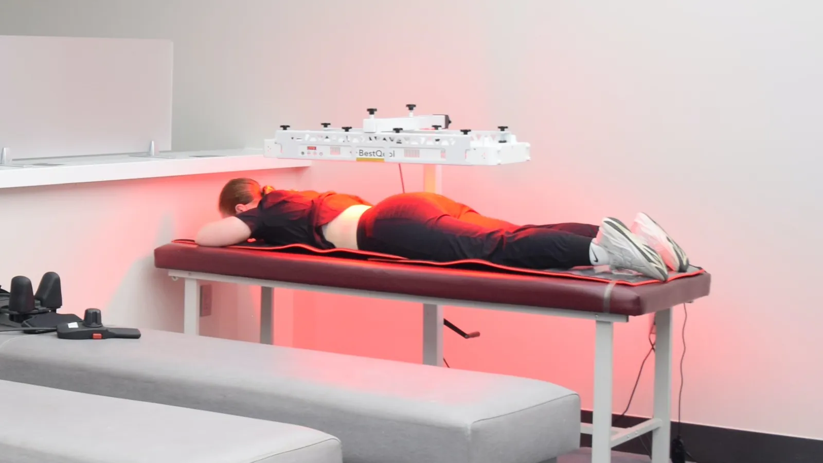
    Patient receiving class 4 photobiomodulation therapy under a red-light panel on a treatment table at Regenerve, 4477 Woodson Rd, St. Louis, MO 63134

    The therapies we add on top

    Once the driver is identified, treatment can be aimed at it. At our St. Louis clinic that toolkit includes class 4 photobiomodulation, class 3B cold laser, whole-body infrared, peripheral and vagal nerve stimulation, nutritional counseling, balance and gait training, and wound care where protective sensation has been lost. Orthobiologic injections including PRP and BMAC are also offered, used as adjuncts inside a plan — never as a substitute for finding the cause. The full list is on our services page, and the reasoning behind the sequence is in the Regenerve Protocol.

    Supplements and complementary approaches

    Some have real randomized evidence — alpha-lipoic acid, acetyl-L-carnitine, palmitoylethanolamide and acupuncture among them — and some widely sold products have failed when properly tested. We grade them one by one in alternative medicine for peripheral neuropathy.

    Hand holding a round mirror to inspect the sole of a foot, the daily self-check recommended when protective sensation is reduced by peripheral neuropathy

    Protecting the feet while you treat them

    When sensation drops below a certain threshold, the foot stops reporting injury. That is what makes neuropathy dangerous rather than merely uncomfortable. A 10-gram monofilament test identifies loss of protective sensation, and anyone who has it needs daily visual foot checks, properly fitted footwear, and prompt attention to any blister, crack or callus — because the usual warning signal is gone.9 Never soak numb feet in water you have not tested with your hand or a thermometer.

    Honest expectations

    Nerve tissue repairs slowly and not all damage is reversible. Progress is measured in reduced pain, better sleep, steadier balance and improved function — usually over months, not weeks. Anyone promising a cure for peripheral neuropathy is telling you something that cannot be supported. What can often be changed is the trajectory, particularly when an active driver is found and treated.

    Being evaluated in St. Louis

    Regenerve is at 4477 Woodson Rd #104, St. Louis, MO 63134, minutes from St. Louis Lambert International Airport, and is led by Dr. Gurpreet Singh Padda, MD, MBA, MHP. The practice serves the St. Louis region, Missouri and Illinois. Electrodiagnostic testing is performed on site, so the evaluation and the results happen in the same place.

    Call or text (314) 886-5902, or email info@regenerve.com. Office hours are Monday through Thursday 9:00 to 16:00 and Friday 9:00 to 13:30. Details are on the contact page and the locations page.

    The clearest place to begin is the Nerve Damage Score, a five-question assessment that produces a large-print report you can bring to any physician.

    Frequently asked questions

    What is the best treatment for peripheral neuropathy in the feet?

    There is no single best treatment, because the right one depends on what is damaging the nerve. Pain medication, topical capsaicin, metabolic correction and nerve-directed therapies all have a place, and the plan should follow the diagnosis rather than precede it. See the Regenerve Protocol.

    What is the best treatment for diabetic neuropathy in the feet?

    There is no single drug that fixes it. In diabetes the nerve damage is driven by the metabolic terrain, so the treatment that changes the nerve is the treatment aimed at that terrain — and nerve fibers can regrow, measured on skin biopsy, though it is work measured in months and no one can promise a cure.

    Pain-directed medication is worth having in the meantime, but it helps not to confuse the two: gabapentinoids, SNRIs and tricyclics treat the pain and not the nerve damage, and the major oral classes cluster tightly together in effect.

    The reason the plan cannot stop at glucose is the evidence itself. A Cochrane review of 17 randomized studies found that tightening glucose control clearly prevents clinical neuropathy in type 1 diabetes, but in type 2 — the population most of our patients belong to — it reduced incidence without reaching statistical significance, while significantly raising the risk of severe hypoglycemia. Glucose is one lever and not the whole plan: lipids, blood pressure, weight and insulin resistance belong in the neuropathy plan rather than to a different doctor. See diabetic peripheral neuropathy treatment in St. Louis.

    Can peripheral neuropathy in the feet be reversed?

    Some of it can. Diet and exercise counseling in patients with impaired glucose tolerance produced measurable regrowth of nerve fibers on skin biopsy after one year. Not all damage is reversible, and no one can promise a cure. See how sugar damages nerves.

    My A1c is normal. Can my neuropathy still be metabolic?

    Yes. In one study of patients with idiopathic neuropathy, 34% had impaired glucose tolerance that a normal fasting glucose and normal HbA1c had missed, and only the two-hour glucose tolerance test revealed it. See how sugar damages nerves.

    Should I be taking gabapentin or pregabalin?

    They are reasonable options with a modest average effect, comparable to SNRIs, tricyclics and sodium channel blockers. They treat the pain and not the nerve damage, so they work best alongside treatment aimed at the cause. See the hidden drivers of peripheral neuropathy.

    Are opioids used for neuropathy in the feet?

    The American Academy of Neurology recommends against using opioids for painful diabetic polyneuropathy. Our practice reduces opioid burden rather than adding to it. See which specialist you should see.

    How long before treatment helps?

    Symptom-directed treatments can work within weeks. Treatment aimed at the underlying metabolic driver is measured in months, because it depends on nerve fibers regrowing. See the Regenerve Protocol.

    Sources

    1. Tesfaye S, et al. Diabetic neuropathies: update on definitions, diagnostic criteria, estimation of severity, and treatments. Diabetes Care. 2010. PMID 20876709
    2. Hicks CW, Wang D, Matsushita K, Windham BG, Selvin E. Peripheral Neuropathy and All-Cause and Cardiovascular Mortality in U.S. Adults: A Prospective Cohort Study. Annals of Internal Medicine. 2021. PMID 33284680
    3. Singleton JR, Smith AG, Bromberg MB. Increased prevalence of impaired glucose tolerance in patients with painful sensory neuropathy. Diabetes Care. 2001. PMID 11473085
    4. Smith AG, Rose K, Singleton JR. Idiopathic neuropathy patients are at high risk for metabolic syndrome. Journal of the Neurological Sciences. 2008. PMID 18606421
    5. Price R, et al. Oral and Topical Treatment of Painful Diabetic Polyneuropathy: Practice Guideline Update Summary. Neurology. 2022. PMID 34965987
    6. Anand P, et al. Reversing painful and non-painful diabetic neuropathy with the capsaicin 8% patch: clinical evidence for pain relief and restoration of function via nerve fiber regeneration. Frontiers in Neurology. 2022. PMID 36388188
    7. Callaghan BC, Little AA, Feldman EL, Hughes RA. Enhanced glucose control for preventing and treating diabetic neuropathy. Cochrane Database of Systematic Reviews. 2012. PMID 22696371
    8. Smith AG, et al. Lifestyle intervention for pre-diabetic neuropathy. Diabetes Care. 2006. PMID 16732011
    9. Boulton AJM, et al. Comprehensive foot examination and risk assessment: a report of the task force of the foot care interest group of the American Diabetes Association. Diabetes Care. 2008. PMID 18663232
  • Which Specialist Should You See for Neuropathy That Has Not Improved?

    Which Specialist Should You See for Neuropathy That Has Not Improved?

    If burning, numbness or nerve pain has not improved after several rounds of treatment, the specialist you need is one who will identify which nerves are affected and what is damaging them — before prescribing anything else. “Neuropathy” names a pattern of symptoms. It is a category, not a diagnosis, and treatment that skips the driver tends to stall.

    That distinction matters because the drivers are so varied. Diabetes is the most common, and 50% to 66% of people with diabetes develop diabetic peripheral neuropathy at some point.1 But a large share of neuropathy is not diabetic at all, and some of it is reversible once the cause is found.

    Why “you have neuropathy” is not an answer

    Being told you have neuropathy tells you your nerves are not working properly. It does not tell you which fibers are involved, how far the damage extends, or why it started.

    Those three answers change the treatment. Small-fiber involvement behaves differently from large-fiber. A compressive problem at a single site is not the same as a symmetrical, length-dependent process. And a nutritional or autoimmune driver calls for something entirely different from a metabolic one.

    What a proper evaluation includes

    Testing that measures nerve function

    Electrodiagnostic testing — EMG and nerve conduction studies — measures how well nerves actually conduct, rather than inferring it from symptoms. Regenerve performs this testing on site, so the evaluation and the results happen in the same place.

    Patient reclined for small-fiber and autonomic nerve testing, with sensor cuffs on both ankles and wrists, at Regenerve, 4477 Woodson Rd, St. Louis, MO 63134

    A search for the driver, not just the label

    This is where most stalled cases are decided. The drivers worth ruling in or out include:

    A fuller account of how these overlap is in the hidden drivers of peripheral neuropathy.

    A treatment plan that follows the finding

    Once the driver is identified, treatment can be aimed at it. The approaches used at Regenerve are described on the services page and include class 4 photobiomodulation, class 3B cold laser, whole-body infrared, and metabolic and nutritional care, alongside correction of whatever the work-up identifies.

    Orthobiologic injections, including PRP and BMAC, are also offered. They are considered as adjuncts within a plan, never as a substitute for identifying the cause.

    Patient receiving class 4 photobiomodulation therapy under a red-light panel on a treatment table at Regenerve, 4477 Woodson Rd, St. Louis, MO 63134

    Questions worth asking any neuropathy clinic

    • Will you test nerve function, or work from symptoms alone?
    • What specific drivers will you rule out, and with which tests?
    • If the cause is metabolic or nutritional, who manages that part?
    • What outcome should I expect, and over what period?
    • What happens if the first approach does not work?

    A clinic that answers these concretely is working from a diagnosis. A clinic that moves straight to a package is not.

    Honest expectations

    Nerve tissue recovers slowly, and not all damage is reversible. Progress is usually measured in reduced pain, better sleep, steadier balance and improved function rather than in a return to how things felt years ago.

    Anyone promising a cure for neuropathy is telling you something that cannot be supported. What can often be changed is the trajectory — particularly when an active driver is found and addressed.

    Being evaluated in St. Louis

    Regenerve is at 4477 Woodson Rd #104, St. Louis, MO 63134, minutes from St. Louis Lambert International Airport, and is led by Dr. Gurpreet Singh Padda, MD, MBA, MHP. The practice serves the St. Louis region, Missouri and Illinois.

    Call or text (314) 886-5902, or email info@regenerve.com. Office hours are Monday through Thursday 9:00 to 16:00 and Friday 9:00 to 13:30. Details are on the contact page and the locations page.

    The clearest place to begin is the Nerve Damage Score, a five-question assessment that produces a large-print report you can bring to any physician. Background on the underlying model is on the science page, and further explanation is on the practice’s YouTube channel, @Regenerve.

    Frequently asked questions

    Which specialist should I see for neuropathy that has not improved?

    One who performs nerve testing on site and investigates the underlying driver rather than treating the label. What that involves is set out on the science page.

    Can neuropathy be reversed?

    Some drivers are correctable and some damage is not. Nutritional and metabolic causes often respond when identified early, as described in functional B12 deficiency and neuropathy. No one can promise a cure.

    My blood sugar is normal. Can it still be metabolic?

    Yes. Nerve injury from glycation can begin below the diabetes threshold, which is why glucose handling is assessed rather than assumed from a single result. See how sugar damages nerves.

    My celiac test was negative but I suspect gluten. Does that rule it out?

    No. Gluten-related nerve injury can occur with a negative celiac panel, which is covered in a negative celiac test does not rule out gluten neuropathy.

    Do you perform nerve testing in the office?

    Yes, electrodiagnostic testing is performed on site. The full list of what is offered is on the services page.

    Do you see patients from Illinois?

    Yes. The practice serves the St. Louis region, Missouri and Illinois, and Dr. Padda is licensed in both states. See the locations page.

    Sources

    1. Bodman MA, Dreyer MA, Varacallo MA. Diabetic Peripheral Neuropathy. StatPearls. NBK442009
  • Sometimes It’s Not Peripheral Neuropathy: Carpal Tunnel, Sciatica, and Double Crush

    Sometimes It’s Not Peripheral Neuropathy: Carpal Tunnel, Sciatica, and Double Crush

    Not every case of numb hands or burning feet is a systemic, metabolic neuropathy. Sometimes the nerve is simply being squeezed — pinched at the wrist, compressed at the spine, or entrapped somewhere along its path. Mechanical compression is a common and often highly treatable cause of nerve symptoms, and distinguishing it from a diffuse neuropathy changes everything about the treatment. This article explains how compression injures nerves, how to recognize the pattern, and why compression and metabolic disease so often team up through a phenomenon called double crush syndrome.

    How compression injures a nerve

    A nerve under sustained mechanical pressure suffers in two main ways. First, compression restricts blood flow to the nerve (ischemia), starving it of oxygen and nutrients. Second, sustained pressure damages the nerve’s insulating myelin at the site of compression (focal demyelination), disrupting the fast, faithful conduction of signals. If the pressure continues, the underlying nerve fibers themselves can be injured. The result is numbness, tingling, pain, and sometimes weakness — but with a crucial difference from metabolic neuropathy: the problem is localized to the compressed nerve, not spread symmetrically across all the longest nerves.

    Common compression syndromes

    Carpal tunnel syndrome is the classic example: the median nerve is compressed as it passes through a tight tunnel at the wrist. It typically causes numbness and tingling in the thumb, index, middle, and part of the ring finger, often worse at night, and sometimes weakness of grip. It is one of the most common nerve disorders and is frequently very treatable.

    Radiculopathy (including sciatica) occurs when a nerve root is compressed as it exits the spine — for example, by a herniated disc or arthritic narrowing. Sciatica is the well-known form: pain, numbness, or weakness radiating from the low back down the leg along the path of the affected nerve root. Because the compression is at the spine, the symptoms follow a specific nerve’s territory rather than a stocking-glove pattern.

    Other entrapments (such as the ulnar nerve at the elbow) follow the same logic: a specific nerve, compressed at a specific spot, producing symptoms in that nerve’s specific distribution.

    Recognizing compression versus diffuse neuropathy

    The pattern is the tell. Metabolic neuropathies (like diabetic neuropathy) are usually symmetric and length-dependent — both feet first, then moving upward. Compression syndromes are usually focal and asymmetric — one nerve, one territory, sometimes provoked by particular positions or activities (typing, a night of a bent wrist, prolonged sitting). Nerve conduction studies and electromyography can localize where along a nerve the problem lies, helping confirm compression and pinpoint its site. Getting this distinction right matters, because a compressed nerve may be relieved by decompression — mechanical or surgical — whereas a metabolic neuropathy needs a metabolic approach.

    Double crush syndrome: when compression and metabolism combine

    Here is one of the most clinically important and underappreciated concepts in nerve medicine. In 1973, Upton and McComas, writing in The Lancet, proposed double crush syndrome: the idea that a nerve compressed at one point becomes more vulnerable to injury at a second point along its length. A single mild compression that might not cause symptoms on its own can become symptomatic when combined with a second insult.

    The insight extends beyond two mechanical compressions. A nerve that is metabolically stressed — by diabetes, by nutritional deficiency, by toxins — is already compromised, and that makes it far more susceptible to symptomatic injury from even modest mechanical compression. This is why so many patients have both: a metabolic neuropathy that has lowered the nerve’s reserve, plus a compression (like carpal tunnel) that pushes it over the threshold into symptoms. Treating only one of the two often leaves the patient frustrated.

    Why a dual approach works best

    The double-crush concept has a direct treatment implication: address both the mechanical and the metabolic contributors. Relieving the compression — through ergonomic changes, splinting, injections, physical therapy, or, when appropriate, decompression procedures — removes the physical insult. Simultaneously supporting the nerve’s metabolic terrain — blood sugar, nutrients, mitochondrial energy, inflammation — raises its resilience so it can tolerate normal life and heal. Neither alone fully solves a double-crush situation; together they can. This dual philosophy is exactly why a thorough evaluation checks for compression even in someone with known metabolic disease, and checks for metabolic drivers even in someone with an obvious entrapment.

    Where this fits

    Mechanical compression is the mechanical category in the three-domain framework of neuropathy drivers — and double crush syndrome is the bridge that explains why the categories so often overlap. A complete workup deliberately looks across all of them, because the most common real-world scenario is not a single cause but a combination, each amplifying the others.

    Frequently asked questions

    How do I know if it’s carpal tunnel or a general neuropathy?

    Carpal tunnel causes symptoms in a specific hand distribution, often worse at night, on one or both sides. Diffuse neuropathy is usually symmetric and starts in the feet. Nerve conduction studies can distinguish and localize the problem.

    Can I have both compression and neuropathy?

    Yes — and it’s common. Double crush syndrome describes how a metabolically stressed nerve becomes more vulnerable to compression, so the two frequently coexist and compound each other.

    Is surgery always needed for compression?

    No. Many compression syndromes respond to conservative measures — splinting, ergonomics, injections, therapy. Decompression is considered when conservative care is insufficient or damage is progressing.

    Why didn’t treating my diabetes fix my hand numbness?

    Because the hand numbness may be a compression (like carpal tunnel), not the diabetic neuropathy — a classic double-crush situation where both the mechanical and metabolic sides need attention.

    Key takeaways

    • Not all nerve pain is metabolic; mechanical compression is common and often treatable.
    • Carpal tunnel and sciatica compress a specific nerve, causing focal, asymmetric symptoms.
    • Compression injures nerves via ischemia and focal demyelination; the pattern distinguishes it from diffuse neuropathy.
    • Double crush syndrome (Upton & McComas, 1973) explains why metabolically stressed nerves are more vulnerable to compression.
    • The best results come from addressing both the mechanical and metabolic contributors together.

    This article is educational and is not a substitute for evaluation, diagnosis, or treatment by a physician. Individual results vary. Take the free Nerve Damage Score or call/text (314) 886-5902.

    References

    1. Upton ARM, McComas AJ. The double crush in nerve-entrapment syndromes. Lancet. 1973;2(7825):359–362.
    2. Padua L, et al. Carpal tunnel syndrome: clinical features, diagnosis, and management. Lancet Neurol. 2016;15:1273–1284.
    3. Wilbourn AJ, Gilliatt RW. Double-crush syndrome: a critical analysis. Neurology. 1997;49:21–29.
    4. Rempel D, et al. Pathophysiology of nerve compression syndromes. J Bone Joint Surg Am. 1999.

    Find out what is driving your nerve pain

    The free, five-question Nerve Damage Score takes about two minutes and tells you which terrain failure is most likely behind your symptoms.

    Get My Free Nerve Damage Score

    Or call or text (314) 886-5902.

  • Mystery Neuropathy and Exhaustion? The Mold and Mycotoxin Question

    Mystery Neuropathy and Exhaustion? The Mold and Mycotoxin Question

    This is a topic that calls for both openness and honesty. Some patients with unexplained neuropathy paired with profound fatigue trace their symptoms to exposure in a water-damaged building, and mold and its toxins are a genuine area of scientific and clinical interest. At the same time, “mold illness” is one of the more contested subjects in medicine, prone to both dismissal on one side and overdiagnosis on the other. This article aims for the middle: what mycotoxins are, the biologically plausible ways they could affect nerves and energy, and how to approach evaluation carefully rather than credulously.

    What mycotoxins are

    Mold is a type of fungus, and some molds produce mycotoxins — toxic compounds that can contaminate the environment, particularly in water-damaged buildings where mold grows on damp materials. Mycotoxins are well established as harmful in certain contexts: contaminated food is a recognized cause of illness in humans and animals, and occupational exposures are studied. The debated question is not whether mycotoxins can be toxic — they can — but how often, and to what degree, indoor mold exposure causes the specific multi-symptom syndromes some patients and practitioners attribute to it.

    The plausible mechanisms

    The mechanisms by which mycotoxins could affect the nervous system are biologically reasonable, which is part of why the topic is taken seriously even amid the controversy.

    Mitochondrial toxicity. Some mycotoxins can impair mitochondrial function in laboratory settings. Since nerves are highly energy-dependent, anything that undermines mitochondrial energy production could, in principle, contribute to nerve dysfunction and to the profound fatigue that often accompanies these presentations — the “drained energy” theme.

    Neuroinflammation. Mycotoxins and mold exposure can provoke immune and inflammatory responses. Chronic neuroinflammation is a recognized contributor to nerve dysfunction, offering another plausible route from exposure to symptoms.

    Oxidative stress. Like other toxins, mycotoxins can increase oxidative stress and deplete antioxidant defenses, adding to the cellular strain on vulnerable tissues.

    These are mechanisms of plausibility, not proof that a given patient’s neuropathy is mold-caused — an important distinction to keep in view.

    The honest state of the evidence

    Transparency matters here. The concept of a chronic multi-system illness from indoor mold exposure (sometimes called “chronic inflammatory response syndrome”) is not universally accepted in mainstream medicine, and some of the testing and treatment marketed for it is unvalidated or commercially driven. Equally, dismissing every patient with real symptoms and a real water-damaged-building exposure does them a disservice. The defensible clinical stance is to take the exposure history and symptoms seriously, evaluate carefully with validated tools, rule in or out the well-established causes of neuropathy first, and avoid both reflexive dismissal and unproven, expensive protocols.

    A careful approach to evaluation

    For someone with unexplained neuropathy, notable fatigue, and a credible exposure to a water-damaged environment, a reasonable, non-credulous approach includes several elements. First, a thorough standard neuropathy workup to identify or exclude the common, well-established drivers — metabolic, nutritional, toxic, autoimmune, and mechanical — because these are more common and more clearly treatable, and a mold attribution should never short-circuit that search. Second, an honest environmental assessment: is there documented water damage and mold in the home or workplace? Removing or remediating a genuinely contaminated environment is sensible regardless of the diagnostic debate. Third, supporting cellular energy and reducing oxidative stress through sound, evidence-based measures — good nutrition, mitochondrial cofactors, sleep, and management of inflammation — which are low-risk and broadly beneficial. And throughout, skepticism toward unvalidated tests and proprietary “detox” protocols that promise to diagnose and cure mold illness, many of which lack rigorous support.

    Where this fits

    Mold and mycotoxins sit within the toxic category of neuropathy drivers, alongside heavy metals, medications, and alcohol. The guiding principle mirrors the heavy-metals discussion: identify and remove genuine environmental exposure, evaluate carefully, and support the body’s resilience — while keeping a clear eye on the difference between plausible mechanism and proven causation, and steering away from the unproven commercial fringe.

    Frequently asked questions

    Can mold really cause neuropathy?

    Mycotoxins are genuinely toxic and can affect mitochondria and inflammation, so a contribution is biologically plausible — but indoor-mold illness is a debated area, and other, better-established causes should be evaluated first.

    That is the subject of Peripheral Neuropathy: The 11 Hidden Drivers Your Doctor Missed.

    Should I get mycotoxin testing?

    Be cautious. Much of the marketed testing is unvalidated and prone to misinterpretation. Focus first on a thorough standard workup and, where relevant, a legitimate environmental assessment of the building.

    There is more on this in Peripheral Neuropathy: The 11 Hidden Drivers Your Doctor Missed.

    Is remediating my home worthwhile?

    If there is documented water damage and mold, addressing it is sensible for general health regardless of the diagnostic debate — removing a real exposure is low-risk and reasonable.

    That is the subject of The Slow Poison in Your Bloodstream: How Heavy Metals Damage Your Nervous System.

    What about mold “detox” protocols?

    Many are unproven and commercially driven. Prioritize evidence-based support (nutrition, sleep, mitochondrial cofactors, inflammation management) and avoid expensive, unvalidated regimens.

    The Slow Poison in Your Bloodstream: How Heavy Metals Damage Your Nervous System goes through it in detail.

    Key takeaways

    • Some molds produce mycotoxins that are genuinely toxic; indoor-mold illness syndromes, however, are medically contested.
    • Plausible mechanisms include mitochondrial toxicity, neuroinflammation, and oxidative stress.
    • The evidence for chronic multi-system mold illness is not settled; avoid both dismissal and overdiagnosis.
    • Evaluate the well-established neuropathy causes first, and assess the environment realistically.
    • Be skeptical of unvalidated testing and proprietary “detox” protocols; favor low-risk, evidence-based support.

    This article is educational and is not a substitute for evaluation, diagnosis, or treatment by a physician. Individual results vary. Take the free Nerve Damage Score or call/text (314) 886-5902.

    References

    1. World Health Organization. WHO Guidelines for Indoor Air Quality: Dampness and Mould. 2009.
    2. Institute of Medicine (US). Damp Indoor Spaces and Health. National Academies Press; 2004.
    3. Empting LD. Neurologic and neuropsychiatric syndrome features of mold and mycotoxin exposure. Toxicol Ind Health. 2009. (Note: represents one side of a contested literature.)
    4. Bennett JW, Klich M. Mycotoxins. Clin Microbiol Rev. 2003;16:497–516.

    Note: this topic is genuinely contested; references are provided to represent the range of evidence, and the article should retain its balanced framing.

    Find out what is driving your nerve pain

    The free, five-question Nerve Damage Score takes about two minutes and tells you which terrain failure is most likely behind your symptoms.

    Get My Free Nerve Damage Score

    Or call or text (314) 886-5902.

  • Neuropathy After Weight-Loss or Gallbladder Surgery? The Deficiency No One Checks

    Neuropathy After Weight-Loss or Gallbladder Surgery? The Deficiency No One Checks

    Some neuropathies trace back to an event that happened years earlier — an operation that solved one problem while quietly setting up another. Surgeries that alter the digestive tract, including bariatric (weight-loss) procedures and gallbladder removal, can impair the absorption of nutrients the nervous system depends on. The nerve damage may appear months or years later, long after anyone is thinking about the surgery, which is exactly why the deficiency so often goes unchecked. This article explains how it happens, which nutrients are at risk, and why lifelong monitoring is the answer.

    How digestive surgery leads to nerve damage

    Nutrient absorption is a choreographed process that depends on specific parts of the digestive tract, specific stomach secretions, and — for fats and fat-soluble vitamins — bile. Surgery that changes this anatomy or physiology can disrupt absorption even when a person eats well.

    Bariatric surgery works partly by reducing how much you eat and, in some procedures, by bypassing sections of the intestine where nutrients are absorbed. It also reduces stomach acid and intrinsic factor, both needed to absorb vitamin B12. The result is a well-documented, long-term risk of multiple nutrient deficiencies.

    Gallbladder removal (cholecystectomy) changes how bile is delivered to the intestine. Because bile is essential for absorbing dietary fat and the fat-soluble vitamins (A, D, E, and K), some people develop impaired fat absorption afterward, which over time can affect nutrients like vitamin E — a deficiency specifically linked to neurological problems.

    The common thread is malabsorption: the food goes in, but the nutrients don’t fully get where they need to go.

    The nutrients at risk — and the nerves they protect

    Several deficiencies after digestive surgery are directly relevant to nerve health.

    Vitamin B12. Highly vulnerable after procedures that reduce stomach acid and intrinsic factor. B12 deficiency causes a characteristic neuropathy and can damage the spinal cord.

    Thiamine (B1). Can become deficient rapidly, especially with vomiting or poor intake after surgery, and thiamine deficiency causes neuropathy and, acutely, can precipitate the neurological emergency Wernicke’s encephalopathy. This is one to catch early.

    Copper. An underrecognized but important one. Copper deficiency — which can follow bariatric surgery (and is worsened by excessive zinc intake) — causes a myeloneuropathy, damaging both peripheral nerves and the spinal cord, sometimes mimicking B12 deficiency. It is frequently missed precisely because few clinicians think to check it.

    Fat-soluble vitamins (especially vitamin E). Impaired fat absorption, more relevant after gallbladder-related changes and malabsorptive procedures, can lead to vitamin E deficiency, which causes a distinct neurological syndrome including neuropathy and balance problems.

    Other B vitamins and minerals. Folate, B6, and others can also run low, compounding the picture.

    Why it’s so often missed

    The delay is the problem. Nutrient stores can take months or years to deplete, so the neuropathy often appears well after the surgery, by which time neither the patient nor a new clinician necessarily connects the two. Copper and vitamin E, in particular, are not on most routine panels, so they go unchecked unless someone specifically thinks of them. A patient can end up labeled with idiopathic neuropathy when the real cause is a treatable, surgery-related deficiency that simply wasn’t on anyone’s radar.

    The answer: lifelong monitoring and repletion

    The solution is straightforward in principle and requires diligence in practice. Anyone who has had bariatric surgery — and those with malabsorption after other digestive procedures — needs lifelong nutritional monitoring, including the nutrients that routine panels skip. That means periodic checks of B12 (functionally, with MMA and homocysteine), thiamine, copper, and the fat-soluble vitamins including vitamin E, along with folate and others as indicated, and repletion tailored to what testing shows.

    Repletion sometimes requires specific forms or routes (for example, higher-dose or non-oral B12) because the absorption problem that caused the deficiency also affects how supplements are absorbed. This is why post-surgical nutritional care is an ongoing medical partnership, not a one-time fix — and when a neuropathy does appear, checking this full panel can reveal a cause that is genuinely correctable.

    Where this fits

    Post-surgical malabsorption is one of the nutritional/metabolic drivers considered in a complete neuropathy workup. Its signature is the combination of an unexplained neuropathy and a history of digestive surgery — a history worth volunteering to any clinician evaluating nerve symptoms, because it points straight at a set of specific, testable deficiencies.

    Frequently asked questions

    I had weight-loss surgery years ago — could that be causing my neuropathy now?

    Yes. Deficiencies can take months to years to produce symptoms, so neuropathy appearing well after surgery is consistent with malabsorption. A targeted nutrient panel can clarify it.

    The mechanism is covered in Nutritional deficiencies beyond B12 that damage peripheral nerves.

    Which deficiencies get missed the most?

    Copper and vitamin E are frequently overlooked because they aren’t on standard panels, yet both cause neurological damage. B12 and thiamine also warrant checking.

    The mechanism is covered in Your “Normal” B12 May Be Lying: The Hidden Deficiency Driving Your Neuropathy.

    I take a multivitamin — isn’t that enough?

    Not always. Malabsorption can outpace a standard multivitamin, and some nutrients need specific forms or doses. Monitoring confirms whether your regimen is actually maintaining adequate levels.

    Your “Normal” B12 May Be Lying: The Hidden Deficiency Driving Your Neuropathy explains what that looks like.

    Does gallbladder removal cause neuropathy?

    It can contribute in some people by impairing fat and fat-soluble-vitamin absorption over time, making vitamin E deficiency worth checking when neuropathy appears.

    This is set out in Nutritional deficiencies beyond B12 that damage peripheral nerves.

    Key takeaways

    • Bariatric and gallbladder surgery can impair nutrient absorption and cause delayed neuropathy.
    • Key nutrients at risk: B12, thiamine, copper, and fat-soluble vitamins (especially vitamin E).
    • Copper and vitamin E deficiencies are commonly missed because they aren’t on routine panels.
    • The delay between surgery and symptoms is why the cause is so often overlooked.
    • Lifelong monitoring and tailored repletion can prevent and reverse these deficiencies.

    This article is educational and is not a substitute for evaluation, diagnosis, or treatment by a physician. Individual results vary. Do not start, stop, or change supplements or medication without consulting your physician. Take the free Nerve Damage Score or call/text (314) 886-5902.

    References

    1. Kumar N. Neurologic complications of bariatric surgery. Continuum (Minneap Minn). 2014.
    2. Jaiser SR, Winston GP. Copper deficiency myelopathy and neuropathy. J Neurol. 2010;257:869–881.
    3. Becker DA, et al. Neurological complications of nutritional deficiency following bariatric surgery. J Obes. 2012.
    4. Kumar N. Nutritional neuropathies (vitamin E, B12, thiamine, copper). Neurol Clin. 2007.

    Find out what is driving your nerve pain

    The free, five-question Nerve Damage Score takes about two minutes and tells you which terrain failure is most likely behind your symptoms.

    Get My Free Nerve Damage Score

    Or call or text (314) 886-5902.

  • Shingles Pain That Won’t Stop? Understanding Postherpetic Neuralgia

    Shingles Pain That Won’t Stop? Understanding Postherpetic Neuralgia

    For most people, shingles is a miserable few weeks and then it’s over. But for a significant minority, the rash heals and the pain doesn’t — it settles into a persistent, often severe nerve pain that can last months or years. This is postherpetic neuralgia (PHN), one of the most common and most distressing forms of neuropathic pain. This article explains why a childhood chickenpox virus can come back decades later to injure nerves, what makes the pain persist, how it is treated, and how it can be prevented.

    A virus that never left

    Shingles is caused by the varicella-zoster virus (VZV) — the same virus that causes chickenpox. After a childhood chickenpox infection, the virus is not eliminated. It retreats into the dorsal root ganglia, the clusters of sensory nerve cell bodies that sit alongside the spinal cord, and lies dormant there, sometimes for decades, held in check by the immune system.

    When immunity wanes — with age, illness, stress, or immune-suppressing conditions and treatments — the virus can reactivate. It travels back down the sensory nerve to the skin, producing the characteristic painful, blistering rash of shingles in a band along the territory of that nerve. But the damage isn’t only skin-deep: the reactivation inflames and injures the nerve and its ganglion along the way.

    Why the pain persists: postherpetic neuralgia

    In postherpetic neuralgia, the nerve injury from the shingles episode leaves the affected sensory pathway damaged and dysregulated. The injured nerves become hyperexcitable and misfire, and the pain-processing system itself can become sensitized, so that even light touch on the healed skin triggers severe pain (a phenomenon called allodynia). The result is persistent burning, stabbing, or electric pain in the area where the rash was, often accompanied by exquisite sensitivity.

    Several factors raise the risk of PHN, most notably older age — the risk climbs substantially with each decade — along with greater severity of the initial shingles episode. This is why prevention and early treatment matter so much.

    Treating postherpetic neuralgia

    PHN is challenging, but there are real tools, and they work best in combination and tailored to the individual.

    Early antiviral treatment of the acute shingles episode (started promptly, within the first days of the rash) can reduce the severity and duration of the outbreak and may lower the risk of persistent pain — one reason to seek care quickly when shingles appears.

    For established PHN, treatments include topical therapies — notably the high-concentration 8% capsaicin patch, which is FDA-approved for postherpetic neuralgia and works by quieting the overactive pain fibers in the affected skin (discussed in detail in the capsaicin article), and lidocaine patches — as well as oral neuropathic-pain medications such as gabapentinoids and certain antidepressants. Because PHN is often localized, the topical, non-systemic options are especially valuable for avoiding whole-body side effects.

    Beyond symptom control, a repair-and-support philosophy addresses the injured nerve terrain itself. The video notes the use of measures aimed at supporting recovery, including cannabinoid-based therapies and glutathione. In the interest of accuracy: cannabinoids have a growing but still-evolving evidence base for neuropathic pain, and glutathione support for nerve recovery is biologically rational but not a proven cure — both belong in the category of adjuncts used within an individualized, physician-guided plan rather than established standalone treatments.

    Prevention: the most important tool

    The single most effective way to deal with postherpetic neuralgia is to prevent the shingles episode that causes it. The recombinant shingles vaccine (Shingrix) is highly effective at preventing shingles and, by extension, PHN, and is recommended for older adults and certain immunocompromised individuals. For anyone in an at-risk group who has not been vaccinated, this is a conversation worth having with a physician — preventing the outbreak is far easier than treating the pain it can leave behind.

    Where this fits

    Postherpetic neuralgia is one of the infection-related drivers of neuropathic pain. Unlike the diffuse, length-dependent neuropathies of metabolic disease, it is typically localized to the nerve territory affected by the shingles outbreak — a distinct pattern that helps identify it and shapes the localized, terrain-supportive approach to treatment.

    Frequently asked questions

    How long does postherpetic neuralgia last?

    It varies widely — from months to years. Some cases resolve gradually; others persist. Older age and a severe initial outbreak increase the likelihood of long-lasting pain.

    Can the capsaicin patch help PHN?

    Yes. The 8% capsaicin patch is FDA-approved for postherpetic neuralgia and can reduce the localized nerve pain by quieting overactive pain fibers; treatment can be repeated.

    Does treating shingles early prevent PHN?

    Prompt antiviral treatment of the acute episode can reduce its severity and may lower the risk of persistent pain, which is why fast care for a shingles rash matters.

    Can I prevent shingles altogether?

    Largely, yes. The recombinant shingles vaccine is highly effective and is recommended for older adults and some others — discuss it with your physician.

    Key takeaways

    • Shingles comes from varicella-zoster virus reactivating from the dorsal root ganglia, injuring the nerve.
    • Postherpetic neuralgia is the persistent nerve pain that can follow, driven by nerve damage and sensitization.
    • Risk rises sharply with age and with a severe initial outbreak.
    • Treatment combines topical options (including the FDA-approved 8% capsaicin patch), oral medications, and terrain support; cannabinoids and glutathione are adjuncts, not proven cures.
    • The shingles vaccine is the most effective way to prevent PHN.

    This article is educational and is not a substitute for evaluation, diagnosis, or treatment by a physician. Individual results vary. Do not start, stop, or change any medication without consulting your physician. Take the free Nerve Damage Score or call/text (314) 886-5902.

    References

    1. Johnson RW, Rice ASC. Postherpetic neuralgia. N Engl J Med. 2014;371:1526–1533.
    2. Gershon AA, et al. Varicella zoster virus infection. Nat Rev Dis Primers. 2015.
    3. Backonja M, et al. NGX-4010 (capsaicin 8% patch) for postherpetic neuralgia: randomized studies. Lancet Neurol. 2008.
    4. Dooling KL, et al. Recommendations of the ACIP for use of recombinant zoster vaccine (Shingrix). MMWR. 2018.

    Find out what is driving your nerve pain

    The free, five-question Nerve Damage Score takes about two minutes and tells you which terrain failure is most likely behind your symptoms.

    Get My Free Nerve Damage Score

    Or call or text (314) 886-5902.