
Neuropathy Resource Library

  • Alcohol and Nerve Damage: Why Your Feet Burn — and How Recovery Actually Works

    Alcohol and Nerve Damage: Why Your Feet Burn — and How Recovery Actually Works

    Alcohol-related nerve damage is common, often underrecognized, and — encouragingly — one of the more recoverable neuropathies when it is addressed properly. It is also a topic that deserves care and directness rather than judgment. This article explains the two distinct ways alcohol injures nerves, why the burning-feet pattern develops, and what genuine recovery requires — including an important safety note about how not to go about stopping.

    A double assault on the nerves

    Alcohol damages peripheral nerves through two mechanisms working at the same time, which is part of why the resulting neuropathy can be significant.

    Direct toxicity. Alcohol and its primary metabolite, acetaldehyde, are directly toxic to nerve tissue. Acetaldehyde is a reactive compound that damages cellular structures and proteins, and chronic exposure injures the nerve fibers themselves and the machinery that keeps them healthy. This toxic effect is thought to contribute to a length-dependent axonal neuropathy — damage that begins at the ends of the longest nerves and works inward, producing the classic burning, tingling, and numbness in the feet.

    Nutritional depletion. Heavy alcohol use depletes the body of essential nutrients, above all thiamine (vitamin B1). Alcohol interferes with thiamine absorption, storage, and activation, and heavy drinking often displaces nutritious food. Because thiamine is critical for the energy metabolism that nerves depend on, its deficiency causes neuropathy in its own right — and thiamine deficiency has other serious neurological consequences as well, including Wernicke’s encephalopathy, a medical emergency. Other B vitamins and nutrients are frequently depleted too.

    So the alcoholic neuropathy that produces burning feet is usually a combination: nerves poisoned directly and starved of the nutrients they need to function and repair.

    Why it often goes unrecognized

    Alcohol-related neuropathy can develop gradually and be attributed to aging, to diabetes (which frequently coexists), or simply dismissed. People may also be reluctant to disclose their drinking, and clinicians may not ask. The result is a treatable, partly reversible neuropathy that goes unaddressed. An honest conversation about alcohol intake is a genuinely important part of an unexplained-neuropathy workup — not to assign blame, but because it points to a cause that can be acted on.

    How recovery actually works

    The good news is that alcoholic neuropathy has real potential for improvement, because both of its drivers can be reversed. Recovery rests on three pillars.

    1. Reducing alcohol exposure. This is the foundation — the direct toxicity cannot heal while it continues. But how this is done matters enormously (see the safety note below).

    2. Repleting nutrients, especially thiamine. Restoring thiamine and other depleted B vitamins gives nerves back the cofactors they need for energy and repair. In the setting of significant deficiency or heavy use, thiamine repletion is often prioritized and, in some clinical situations, given before glucose to avoid precipitating harm — a detail that underscores why this should be medically guided.

    3. Repairing the terrain. Beyond stopping the insult and replacing nutrients, the injured nerve benefits from the same supportive measures any recovering nerve needs — attention to mitochondrial function, inflammation, and overall metabolic health. As nerve tissue repairs slowly, improvement unfolds over months.

    An important safety note

    Here is a crucial caution that a responsible article must include. For a person who is physically dependent on alcohol, stopping abruptly can be dangerous — alcohol withdrawal can cause seizures and a life-threatening condition called delirium tremens. Reducing or stopping alcohol in the setting of dependence should be done with medical support, which can make the process both safer and more successful. This is not a reason to keep drinking; it is a reason to get help doing it safely. If alcohol use is significant, the first step is a conversation with a physician about a supported plan, not going cold turkey alone.

    Help is available, and seeking it is a sign of strength, not weakness. Recovery from both the dependence and the neuropathy is genuinely possible.

    Where this fits

    Alcohol is one of the toxic drivers evaluated in a complete neuropathy assessment. It frequently overlaps with nutritional deficiency and with diabetes, so identifying it is part of assembling the full picture — and it is one of the more rewarding drivers to address, because meaningful recovery is often achievable.

    Frequently asked questions

    Can alcoholic neuropathy be reversed?

    It has real potential to improve when alcohol exposure is reduced and nutrients — especially thiamine — are replaced, though recovery is gradual and may be partial with advanced damage.

    How much alcohol causes neuropathy?

    There’s no single threshold; risk rises with the amount and duration of heavy use, and it’s compounded by poor nutrition. Coexisting diabetes lowers the margin further.

    Should I just quit cold turkey?

    If you drink heavily or are dependent, no — abrupt cessation can trigger dangerous withdrawal. Reduce or stop with medical support to do it safely.

    Which nutrient matters most?

    Thiamine (B1) is central, but other B vitamins and nutrients are commonly depleted too, so repletion is usually broader than thiamine alone.

    Key takeaways

    • Alcohol injures nerves two ways at once: direct toxicity (acetaldehyde) and thiamine/nutrient depletion.
    • The result is typically a length-dependent neuropathy with burning, tingling, and numb feet.
    • Recovery rests on reducing exposure, repleting thiamine and other nutrients, and supporting nerve repair.
    • Improvement is real but gradual; earlier action preserves more function.
    • In dependence, never stop abruptly — withdrawal can be dangerous; stop with medical support.

    This article is educational and is not a substitute for evaluation, diagnosis, or treatment by a physician. Individual results vary. If you drink heavily, do not stop abruptly without medical guidance. Take the free Nerve Damage Score or call/text (314) 886-5902. If you need support for alcohol use, help is available — talk with a physician or call the SAMHSA National Helpline at 1-800-662-4357.

    References

    1. Chopra K, Tiwari V. Alcoholic neuropathy: possible mechanisms and future treatment possibilities. Br J Clin Pharmacol. 2012;73(3):348–362.
    2. Koike H, et al. Alcoholic neuropathy. Curr Opin Neurol / Muscle Nerve.
    3. Sechi G, Serra A. Wernicke’s encephalopathy: new clinical settings and recent advances. Lancet Neurol. 2007;6:442–455.

    Find out what is driving your nerve pain

    The free, five-question Nerve Damage Score takes about two minutes and tells you which terrain failure is most likely behind your symptoms.

    Get My Free Nerve Damage Score

    Or call or text (314) 886-5902.

  • The Cruel Irony: How Statins and Metformin Can Affect Nerve Health

    The Cruel Irony: How Statins and Metformin Can Affect Nerve Health

    There is an uncomfortable irony at the heart of this topic: two of the most widely prescribed medications in the world — statins for cholesterol and metformin for diabetes — can, in some people, quietly deplete the very nutrients that nerves depend on. This does not mean these drugs are villains; they prevent heart attacks, strokes, and the progression of diabetes, and for most people the benefits are substantial. The point of this article is not to frighten anyone off necessary treatment, but to explain a real, manageable interaction so that the nutrients can be monitored and replaced. The answer here is repletion and monitoring, not abandoning needed medication.

    Metformin and vitamin B12

    Metformin is a cornerstone of diabetes care, and a good one. But a well-documented side effect of long-term use is impaired absorption of vitamin B12. Over years, this can produce a functional or overt B12 deficiency — and because B12 deficiency itself causes peripheral neuropathy, the result can be a patient whose diabetes drug is contributing to the very nerve symptoms being blamed entirely on their diabetes.

    This is one of the clearer, better-studied drug–nutrient interactions, and it has a clean solution: periodic monitoring of B12 status in people on long-term metformin, ideally using functional markers like methylmalonic acid and homocysteine rather than serum B12 alone (as discussed in the B12 article), and repletion when needed. Crucially, this is done without stopping metformin — the drug continues to do its job while the B12 is replaced.

    Statins and CoQ10

    Statins lower cholesterol by inhibiting an enzyme (HMG-CoA reductase) early in the cholesterol synthesis pathway. That same pathway also produces coenzyme Q10 (CoQ10), a molecule essential for mitochondrial energy production. By design, statins can therefore lower CoQ10 levels — and since nerves are metabolically demanding, the theoretical concern is that reduced CoQ10 could affect nerve energy metabolism.

    Honesty is important here, because this is a more debated area than the metformin–B12 link. The CoQ10-depletion mechanism is real. Whether statins meaningfully cause peripheral neuropathy has been studied and remains genuinely contested — some observational studies have suggested an association, while others have not confirmed a clear causal link, and any absolute risk appears small. The most defensible position is measured: statin-associated muscle symptoms are well recognized, a neuropathy association is possible but not firmly established, and the benefits of statins for cardiovascular protection are strong and well proven.

    The sensible framework: don’t stop — monitor and support

    Put together, these interactions call for a specific, non-alarmist approach.

    First, do not stop a statin or metformin on your own. Discontinuing a needed cardiovascular or diabetes medication carries real, sometimes serious, risks that typically outweigh the nutrient concerns.

    Second, monitor. In people on long-term metformin, check B12 status functionally and periodically. In people on statins with new muscle or nerve symptoms, discuss them with the prescribing clinician.

    Third, replete intelligently. Where B12 is low, replace it. Where CoQ10 supplementation is being considered for statin-related symptoms, that is a reasonable, low-risk conversation to have with a physician, recognizing that the evidence for benefit is mixed.

    Fourth, look at the whole picture. A person with diabetes on metformin and a statin may have several simultaneous contributors to their neuropathy — the diabetes itself, a metformin-related B12 deficiency, and possibly others. Sorting out how much each contributes is exactly the work of a root-cause evaluation.

    Other medications worth knowing about

    Metformin and statins are the headliners, but they are not alone. Certain chemotherapy agents are directly neurotoxic and a well-known cause of neuropathy; some antibiotics (including certain fluoroquinolones and long-term use of others) and a handful of other drugs can affect nerves as well. The recurring principle applies: the goal is informed monitoring and, where possible, mitigation — in partnership with the prescriber — rather than reflexive discontinuation.

    Frequently asked questions

    Should I stop my metformin or statin if I have neuropathy?

    No. Never stop these on your own — the risks of doing so are significant. Instead, ask your physician to check the relevant nutrient status and evaluate your symptoms.

    Does metformin definitely cause B12 deficiency?

    Long-term metformin use is a well-documented cause of impaired B12 absorption in a meaningful subset of users. It is manageable by monitoring and repletion while continuing the drug.

    Do statins cause neuropathy?

    The CoQ10-depletion mechanism is real, but whether statins meaningfully cause peripheral neuropathy is debated and any risk appears small. Muscle symptoms are the better-recognized statin side effect. Discuss new symptoms with your prescriber.

    Is it safe to take CoQ10 with a statin?

    CoQ10 is generally low-risk, and some people take it for statin-related symptoms, though the evidence for benefit is mixed. Discuss it with your physician.

    Key takeaways

    • Metformin can impair B12 absorption over time, contributing to neuropathy — a well-documented, manageable interaction.
    • Statins can lower CoQ10; a neuropathy link is possible but debated, and any risk appears small.
    • The answer is monitoring and repletion, never stopping needed medication on your own.
    • Functional B12 testing catches deficiencies a standard test misses in metformin users.
    • Multiple contributors often coexist, which is why the full picture matters.

    This article is educational and is not a substitute for evaluation, diagnosis, or treatment by a physician. Individual results vary. Do not start, stop, or change any medication without consulting your physician. Take the free Nerve Damage Score or call/text (314) 886-5902.

    References

    1. Aroda VR, Edelstein SL, Goldberg RB, et al. Long-term metformin use and vitamin B12 deficiency in the Diabetes Prevention Program Outcomes Study. J Clin Endocrinol Metab. 2016;101(4):1754–1761.
    2. Infante M, et al. Metformin, vitamin B12 deficiency and peripheral neuropathy (review). Endocrine / review.
    3. Marcoff L, Thompson PD. The role of coenzyme Q10 in statin-associated myopathy. J Am Coll Cardiol. 2007;49:2231–2237.
    4. Emad M, et al. Statins and peripheral neuropathy — evidence and controversy (review).

    Find out what is driving your nerve pain

    The free, five-question Nerve Damage Score takes about two minutes and tells you which terrain failure is most likely behind your symptoms.

    Get My Free Nerve Damage Score

    Or call or text (314) 886-5902.

  • The Slow Poison in Your Bloodstream: How Heavy Metals Damage Your Nervous System

    The Slow Poison in Your Bloodstream: How Heavy Metals Damage Your Nervous System

    Some causes of neuropathy announce themselves. Heavy-metal toxicity does the opposite: it accumulates quietly, often over years, from sources a person may never connect to their symptoms — old plumbing, contaminated water, certain occupations, some imported products. By the time nerve damage appears, the exposure may be long-standing. This article explains how metals like lead, arsenic, and mercury injure the nervous system, when to suspect them, and — just as importantly — why the popular world of aggressive “detox” can do more harm than the metals themselves.

    Why metals are so toxic to nerves

    Heavy metals damage nerves through a few overlapping mechanisms. They disrupt essential enzymes by binding to sulfur-containing sites the enzymes need to function, effectively jamming the machinery of the cell. They deplete glutathione, the body’s master antioxidant, leaving nerves defenseless against oxidative stress. And they interfere with mitochondrial energy production, starving the energy-hungry nerve of the fuel it needs. The nervous system, with its high metabolic demand and long, vulnerable fibers, is among the tissues most sensitive to these insults.

    The main offenders

    Lead. A classic cause of peripheral neuropathy, historically associated with a motor-predominant pattern (weakness, sometimes wrist drop). Exposure can come from old lead paint and pipes, certain occupations (battery work, smelting, construction, radiator repair), contaminated soil, and some traditional remedies and imported goods.

    Arsenic. Causes a painful sensory neuropathy that can resemble other length-dependent neuropathies, often with characteristic skin changes. Exposure sources include contaminated groundwater (a significant issue in some regions), certain pesticides, and industrial processes.

    Mercury. Affects both the peripheral nerves and the central nervous system. Sources include certain fish (methylmercury), some industrial exposures, and older dental or medical materials.

    Thallium and others. Less common but notable causes of severe neuropathy, sometimes with hair loss as a clue.

    When to suspect metal toxicity

    Heavy-metal neuropathy is worth considering when there is a plausible exposure history — an occupation, hobby, water source, or product that could carry the metal — particularly when the neuropathy is otherwise unexplained, is progressing, or is accompanied by systemic clues (abdominal symptoms, anemia, skin or nail changes, cognitive complaints). The history is the single most important guide; without a reason to suspect exposure, indiscriminate testing tends to generate more confusion than clarity.

    Testing done right

    When exposure is plausible, testing should be targeted and appropriate to the specific metal. Different metals require different specimens and timing — for instance, some are best assessed in blood for recent exposure and others in urine or with provoked testing under careful supervision. This is a domain where testing should be interpreted by a clinician familiar with toxicology, because reference ranges, specimen types, and the meaning of results all depend on the metal and the exposure timeline.

    The detox trap

    Here is the part that deserves a blunt warning. The internet is full of “heavy-metal detox” protocols — supplements, cleanses, and chelation regimens marketed for everything from fatigue to neuropathy. Many are unproven, some are useless, and a few are genuinely dangerous. Aggressive or improperly supervised chelation can redistribute metals to sensitive tissues, cause serious electrolyte and kidney problems, and has been linked to harm. Chelation therapy has legitimate, specific medical indications for confirmed significant toxicity — but it is a medical treatment with real risks, not a wellness product, and it should only ever be done under qualified medical supervision after appropriate testing confirms a treatable burden.

    The responsible approach is the opposite of a generic cleanse: identify and remove the source of exposure, confirm the specific metal with proper testing, support the body’s own detoxification pathways (including glutathione status) sensibly, and reserve chelation for the specific, confirmed situations where medical evidence supports it.

    Where this fits in the bigger picture

    Heavy metals are one of the toxic drivers evaluated in a complete root-cause neuropathy workup, alongside medications, alcohol, and mold. As with the others, the value of identifying a metal contribution is that removing the exposure can halt ongoing damage — but it has to be identified correctly first, which is why history-guided, targeted evaluation matters so much here.

    Frequently asked questions

    Could heavy metals be causing my neuropathy without my knowing?

    It’s possible if you have a genuine exposure source. Because accumulation is slow and silent, the link is easy to miss — which is why exposure history is key.

    Should I get a heavy-metal panel just to check?

    Testing is most useful when guided by a plausible exposure. Untargeted “panels,” especially provoked urine tests marketed online, are prone to misinterpretation. Discuss appropriate testing with a clinician.

    Are detox supplements or cleanses a good idea?

    Generally no. Many are unproven, and aggressive chelation carries real risks. Legitimate treatment for confirmed toxicity is a supervised medical process, not a store-bought cleanse.

    What’s the most important first step?

    Finding and removing the source of exposure. No treatment works while exposure continues.

    Key takeaways

    • Heavy metals injure nerves by disrupting enzymes, depleting glutathione, and impairing mitochondria.
    • Lead, arsenic, and mercury are the main offenders, each with characteristic patterns and sources.
    • Suspicion should be driven by a real exposure history; testing must be targeted and expertly interpreted.
    • Removing the source is the essential first step.
    • Unproven “detox” and unsupervised chelation can be harmful; chelation is a supervised medical treatment for confirmed toxicity only.

    This article is educational and is not a substitute for evaluation, diagnosis, or treatment by a physician. Individual results vary. Do not attempt detoxification or chelation without qualified medical supervision. Take the free Nerve Damage Score or call/text (314) 886-5902.

    References

    1. Agency for Toxic Substances and Disease Registry (ATSDR). Toxicological Profiles: Lead, Arsenic, Mercury.
    2. Thomson RM, Parry GJ. Neuropathies associated with excessive exposure to lead. Muscle Nerve. 2006.
    3. Rao DG, et al. Arsenic-induced peripheral neuropathy. Pract Neurol / review.
    4. Kern JK, et al. Cautions regarding chelation and unproven detoxification; glutathione and heavy-metal toxicity (review).

    Find out what is driving your nerve pain

    The free, five-question Nerve Damage Score takes about two minutes and tells you which terrain failure is most likely behind your symptoms.

    Get My Free Nerve Damage Score

    Or call or text (314) 886-5902.

  • Caught in the Crossfire: How Lupus and Rheumatoid Arthritis Attack Your Nerves

    Caught in the Crossfire: How Lupus and Rheumatoid Arthritis Attack Your Nerves

    When the immune system is dysregulated, nerves are rarely the intended target — but they are frequently the casualties. Autoimmune diseases such as lupus, rheumatoid arthritis, and Sjögren’s syndrome can produce peripheral neuropathy as collateral damage from a body-wide inflammatory process. This article explains the two main routes by which autoimmunity injures nerves, why the resulting neuropathy can take several distinct forms, and why effective treatment must address both the systemic disease and the nerve.

    Nerves as collateral damage

    In an autoimmune disease, the immune system loses the ability to distinguish self from non-self and begins attacking the body’s own tissues. In lupus (systemic lupus erythematosus) and rheumatoid arthritis, the headline targets are joints, skin, kidneys, and other organs — but the same inflammatory machinery can reach the peripheral nerves. Understanding neuropathy in this setting means understanding that the nerve damage is a downstream consequence of a systemic process, which is why simply treating the nerve is not enough.

    Route one: vasculitis — choking the nerve’s blood supply

    Peripheral nerves depend on a network of tiny blood vessels, the vasa nervorum, for their oxygen and nutrients. In autoimmune disease, these vessels can become inflamed — a process called vasculitis. Inflammation thickens and occludes the vessel walls, cutting off blood flow and starving segments of nerve of oxygen (ischemia). The nerve tissue downstream is injured or dies.

    Vasculitic neuropathy often produces a distinctive pattern called mononeuritis multiplex, in which several individual nerves are damaged in a patchy, asymmetric fashion — for example, a wrist drop on one side and a foot drop on the other, rather than the symmetric stocking-glove pattern typical of diabetic neuropathy. This asymmetry is an important clue that points toward a vasculitic, autoimmune cause. Vasculitic neuropathy can progress quickly and is considered a situation that warrants prompt evaluation.

    Route two: direct antibody attack

    The second route is more direct. The autoantibodies and immune complexes generated in autoimmune disease can bind to and attack nerve components themselves, including the myelin sheath and the nerve fibers. Sjögren’s syndrome is particularly associated with sensory neuropathies — including a sensory ganglionopathy in which the sensory nerve cell bodies themselves are targeted — which can cause numbness, imbalance, and painful burning sensations. Different autoimmune conditions favor different antibody targets, which is part of why autoimmune neuropathy is not a single entity but a family of presentations.

    Recognizing the pattern

    Several features raise suspicion that a neuropathy is autoimmune in origin: an asymmetric or patchy distribution (mononeuritis multiplex), a relatively rapid onset, the presence of systemic symptoms (joint pain, rash, dry eyes and mouth, fatigue), and a known or suspected autoimmune diagnosis. When these are present, the workup expands to include autoimmune serologies and, in some cases, nerve conduction studies or nerve biopsy to characterize the process.

    Why treatment must be two-pronged

    This is the central clinical message. Because the neuropathy is driven by a systemic autoimmune process, controlling that process is essential — typically the domain of rheumatologic care, using immune-modulating treatment to quiet the underlying disease. But calming the systemic inflammation does not automatically repair nerves that have already been injured. The nerve itself also needs support: pain management, protection of remaining function, and attention to the same terrain factors — circulation, nutrients, mitochondrial energy — that any injured nerve requires to recover.

    A terrain-focused clinic works alongside a patient’s rheumatologist rather than in place of them: the rheumatologist targets the fire, and the nerve-focused care supports the tissue caught in it. Neither alone is sufficient.

    The overlap with other drivers

    Autoimmune disease also raises the stakes on other neuropathy drivers. Systemic inflammation, certain immune-modulating medications, and reduced activity can all compound nutritional and metabolic contributors, so a complete evaluation still checks the full range of causes rather than stopping at the autoimmune label.

    Frequently asked questions

    How is autoimmune neuropathy different from diabetic neuropathy?

    Diabetic neuropathy is usually symmetric and starts in the feet. Autoimmune neuropathy is often asymmetric and patchy (mononeuritis multiplex) and tends to accompany other systemic symptoms.

    If I control my lupus or RA, will my neuropathy go away?

    Controlling the underlying disease is essential to stop ongoing damage, but nerves already injured need separate support and recover slowly. The two goals go together.

    Can neuropathy be the first sign of an autoimmune disease?

    Sometimes. An unexplained, especially asymmetric, neuropathy can prompt discovery of an underlying autoimmune condition, which is why the workup includes autoimmune testing.

    Should I stop my immune medications if I develop neuropathy?

    No — never adjust these on your own. Some are treating the very process damaging your nerves. Any change is made with your rheumatologist and physician.

    Key takeaways

    • Lupus, RA, and Sjögren’s can damage nerves as collateral damage from systemic autoimmunity.
    • Vasculitis starves nerves of blood, often causing asymmetric mononeuritis multiplex.
    • Direct antibody attack targets myelin and nerve fibers; Sjögren’s favors sensory neuropathies.
    • Asymmetry, rapid onset, and systemic symptoms are clues to an autoimmune cause.
    • Treatment must control the systemic disease and support the injured nerve.

    This article is educational and is not a substitute for evaluation, diagnosis, or treatment by a physician. Individual results vary. Do not start, stop, or change any medication without consulting your physician. Take the free Nerve Damage Score or call/text (314) 886-5902.

    References

    1. Collins MP, Hadden RD. The nonsystemic vasculitic neuropathies. Nat Rev Neurol. 2017;13:302–316.
    2. Mori K, et al. The wide spectrum of clinical manifestations in Sjögren’s syndrome–associated neuropathy. Brain. 2005;128:2518–2534.
    3. Oomatia A, et al. Peripheral neuropathies in systemic lupus erythematosus. Arthritis Rheumatol. 2014.
    4. Gwathmey KG, et al. Vasculitic neuropathies. Lancet Neurol. 2014;13:67–82.

    Find out what is driving your nerve pain

    The free, five-question Nerve Damage Score takes about two minutes and tells you which terrain failure is most likely behind your symptoms.

    Get My Free Nerve Damage Score

    Or call or text (314) 886-5902.

  • Negative Celiac Test but Burning Feet? The Gluten–Neuropathy Link Doctors Miss

    Negative Celiac Test but Burning Feet? The Gluten–Neuropathy Link Doctors Miss

    You did the responsible thing. You mentioned your burning feet, someone ordered a celiac panel, it came back negative, and gluten was crossed off the list. But for a large number of people with gluten-related nerve damage, that negative result is exactly where the diagnosis goes wrong. The celiac test is answering a different question than the one your nerves are asking. This article explains why standard celiac screening misses gluten neuropathy, and what testing actually reveals it.

    The celiac test answers the wrong question

    Celiac disease is defined by an immune attack on the small intestine. Its screening tools are built to detect that — chiefly antibodies to tissue transglutaminase-2 (tTG-2), the transglutaminase isoform concentrated in the gut, confirmed by intestinal biopsy showing damage to the villi.

    Gluten-related neurological disease is a different manifestation of gluten sensitivity. The immune response targets the nervous system, and the gut may be entirely spared. A patient can therefore have a pristine celiac panel and normal intestinal biopsy while producing the antibodies that are injuring their peripheral nerves. The test isn’t broken — it’s simply looking in the wrong place.

    What the immune system actually targets in the nerves

    As covered in the companion article on the gluten–neuropathy connection, the mechanism is molecular mimicry. Two targets are especially relevant to a nerve-focused workup.

    Transglutaminase-6 (TG6). This isoform is expressed in nervous tissue, and antibodies against it — described by Dr. Marios Hadjivassiliou’s group — mark the neurological form of gluten sensitivity. TG6 antibodies can be present when the gut-focused tTG-2 antibodies are absent, which is precisely why a celiac screen can miss neurological disease.

    Anti-myelin and related antibodies. The immune response can also target components of the myelin sheath that insulates nerves, contributing to the sensory neuropathy and, when the cerebellum is involved, the ataxia seen in gluten-related neurological disease.

    The genetics: HLA-DQ2 and HLA-DQ8

    Gluten sensitivity has a strong genetic underpinning. The great majority of people who react to gluten — whether the celiac or the neurological form — carry one of two immune-system genetic markers, HLA-DQ2 or HLA-DQ8. Testing for these haplotypes is informative in a different way than antibody testing: a negative HLA-DQ2/DQ8 result makes gluten-driven disease very unlikely and can effectively rule it out, while a positive result establishes susceptibility (though not everyone who is susceptible develops disease). This makes HLA typing a useful gatekeeping test in an unexplained neuropathy workup.

    Building a proper gluten-neuropathy panel

    For someone with an unexplained sensory neuropathy — especially with any balance disturbance — a more complete evaluation than a routine celiac screen includes HLA-DQ2/DQ8 genetic typing, an expanded gluten-related antibody panel (including TG6 where available, alongside the conventional celiac antibodies), and a clinical assessment for the length-dependent sensory pattern and any cerebellar signs. The aim is to catch the neurological form that the standard screen is not designed to see.

    An important practical caveat: antibody testing is most meaningful while gluten is still in the diet. If a patient has already gone gluten-free, antibody levels may fall and produce falsely reassuring results — something to plan around with a clinician before testing.

    Why it’s worth the effort

    Chasing down a gluten contribution is worthwhile because, unlike many drivers of neuropathy, it is entirely removable — the trigger is dietary. When gluten-related neurological disease is confirmed and strict elimination is undertaken early, symptoms can stabilize and sometimes improve. The tragedy of the missed diagnosis is that the person keeps eating the trigger for years, attributing their progressive nerve damage to bad luck or aging, when a change on the plate could have changed the trajectory.

    This is one driver among several

    Gluten sensitivity is one possible contributor evaluated within a broader root-cause investigation that also weighs metabolic, toxic, and mechanical causes. Because multiple drivers frequently coexist, ruling gluten in or out is part of assembling the complete picture rather than a search for a single culprit.

    Frequently asked questions

    My celiac test was negative — can gluten still be damaging my nerves?

    Yes. Celiac screening detects the intestinal form of gluten disease. The neurological form can occur with a negative celiac panel and normal gut biopsy, driven by antibodies like TG6.

    What should I ask to be tested for instead?

    Consider HLA-DQ2/DQ8 genetic typing and an expanded gluten-related antibody panel including transglutaminase-6 where available, ideally while still eating gluten.

    If I already went gluten-free, will testing still work?

    Antibody levels can fall after gluten removal, which may make results falsely negative. Discuss timing with your physician before testing.

    Does a positive HLA-DQ2/DQ8 mean I definitely have gluten neuropathy?

    No — it establishes susceptibility, not disease. It’s most useful in the reverse: a negative result makes gluten-driven disease very unlikely.

    Key takeaways

    • Standard celiac tests detect intestinal disease, not the neurological form of gluten sensitivity.
    • Transglutaminase-6 (TG6) antibodies mark the neural attack and can be present with a negative celiac panel.
    • HLA-DQ2/DQ8 typing is a useful gatekeeper: a negative result largely rules gluten out.
    • Test while still eating gluten, since prior elimination can produce falsely negative antibodies.
    • Confirmed gluten neuropathy is uniquely treatable, because the trigger is removable.

    This article is educational and is not a substitute for evaluation, diagnosis, or treatment by a physician. Individual results vary. Do not make major dietary or medication changes without consulting your physician. Take the free Nerve Damage Score or call/text (314) 886-5902.

    References

    1. Hadjivassiliou M, et al. Transglutaminase-6 antibodies in the diagnosis of gluten ataxia. Neurology. 2013;80:1740–1745.
    2. Hadjivassiliou M, Sanders DS, Grünewald RA, et al. Gluten sensitivity: from gut to brain. Lancet Neurol. 2010;9:318–330.
    3. Hadjivassiliou M, et al. Gluten-related neurological dysfunction and HLA associations.
    4. Volta U, et al. Serological tests in gluten-related disorders. Cell Mol Immunol / review.

    Find out what is driving your nerve pain

    The free, five-question Nerve Damage Score takes about two minutes and tells you which terrain failure is most likely behind your symptoms.

    Get My Free Nerve Damage Score

    Or call or text (314) 886-5902.

  • Your “Normal” B12 May Be Lying: The Hidden Deficiency Driving Your Neuropathy

    Your “Normal” B12 May Be Lying: The Hidden Deficiency Driving Your Neuropathy

    One of the most treatable causes of peripheral neuropathy is also one of the most frequently missed — not because doctors don’t test for it, but because the usual test can read “normal” while the deficiency is doing real damage at the cellular level. This article explains why a serum B12 result can be misleading, which functional markers reveal what it hides, and the full roster of nutrients your nerves depend on to function and to heal.

    Why a “normal” B12 can be wrong

    The standard test measures the total amount of vitamin B12 circulating in your blood. The problem is that this number does not necessarily reflect how much B12 is actually available and working inside your cells. A substantial portion of measured B12 is bound to a protein that cannot deliver it to tissues, so the total can look adequate while the metabolically active supply is low. Reference ranges also vary, and results in the low-normal zone are commonly reported as fine even when a patient is symptomatic.

    The consequence is a patient with the classic tingling, numbness, and imbalance of a B12-related neuropathy being told their levels are “normal,” while the underlying deficiency quietly progresses. Untreated, B12 deficiency can damage not only peripheral nerves but the spinal cord itself — and while early damage is reversible, advanced damage may not be.

    The functional markers that tell the truth: MMA and homocysteine

    To see what serum B12 hides, clinicians use functional markers — substances that accumulate when B12 is inadequate at the cellular level, regardless of what the total blood level says.

    Methylmalonic acid (MMA). B12 is a required cofactor for the enzyme that processes methylmalonyl-CoA. When B12 is functionally deficient inside cells, this reaction stalls and MMA builds up. An elevated MMA is a sensitive and relatively specific sign of true B12 deficiency at the tissue level.

    Homocysteine. B12 (along with folate and B6) is needed to convert homocysteine into methionine. When B12 is functionally low, homocysteine rises. Elevated homocysteine is less specific than MMA — it can also reflect folate or B6 deficiency — but together the two markers give a far more accurate picture than serum B12 alone.

    Checking MMA and homocysteine can catch a functional deficiency that a normal B12 would have masked, allowing correction before permanent nerve or spinal-cord injury occurs.

    Common reasons B12 runs low

    Functional B12 deficiency is especially worth checking in several situations. Metformin, one of the most-prescribed diabetes medications, interferes with B12 absorption and can produce deficiency over years of use — a cruel irony in patients whose diabetes is already threatening their nerves. Acid-suppressing drugs (PPIs and H2 blockers) reduce the stomach acid needed to liberate B12 from food. Age reduces absorption, as can autoimmune (pernicious) causes, gastrointestinal surgery, and strict plant-based diets. Anyone with unexplained neuropathy and any of these risk factors deserves functional testing.

    B12 is not alone: the nutrient team your nerves need

    B12 gets the headlines, but nerves depend on a whole team of cofactors, and deficiency of any one can impair function or healing.

    Thiamine (B1). Essential for glucose metabolism and mitochondrial energy production. Thiamine deficiency directly causes neuropathy, and it is often depleted in diabetes and in heavy alcohol use. The work of Dr. Hammes and colleagues highlighted how thiamine-dependent pathways protect against the biochemical damage of high blood sugar — part of the rationale for the thiamine derivative benfotiamine.

    Vitamin B6. Required for nerve function and neurotransmitter synthesis — but with an important caveat: both deficiency and excess can cause neuropathy, so B6 supplementation should be measured and monitored, not open-ended.

    Magnesium. A cofactor in hundreds of enzymatic reactions, including those governing nerve excitability and mitochondrial energy. Deficiency contributes to nerve hyperexcitability and is common in diabetes.

    Omega-3 fatty acids. Structural components of nerve membranes with anti-inflammatory effects; adequate intake supports the membrane integrity and the inflammatory balance that healthy nerves require.

    The mitochondrial common thread

    Notice the recurring theme: nearly every one of these nutrients feeds into mitochondrial energy production. A nerve is an energy-intensive structure, and it fails when its power supply falters. This is why correcting deficiencies is not simply “topping up vitamins” — it is restoring the cell’s ability to make energy, repair itself, and maintain its signaling. Functional nutrient testing, and correction guided by that testing, is a foundational part of a root-cause neuropathy plan rather than an afterthought.

    What to do with this information

    The practical message is to test properly and correct precisely. That means functional markers (MMA, homocysteine) rather than serum B12 alone, evaluation of thiamine and magnesium status, a review of medications that deplete these nutrients (without stopping needed prescriptions on your own), and repletion tailored to what the testing shows. Because some deficiencies (like B6) can harm in excess, and because absorption problems may require specific forms or routes of supplementation, this is best done under medical guidance.

    Frequently asked questions

    My B12 came back normal — could it still be my problem?

    Yes. Total serum B12 can read normal while the active, cellular supply is low. MMA and homocysteine testing can reveal a functional deficiency the standard test misses.

    See Peripheral Neuropathy: The 11 Hidden Drivers Your Doctor Missed.

    Does metformin cause B12 deficiency?

    It can, over time, by impairing absorption. If you take metformin and have neuropathy symptoms, functional B12 testing is reasonable — but do not stop metformin on your own.

    There is more on this in The Cruel Irony: How Statins and Metformin Can Affect Nerve Health.

    Can I just take high-dose B vitamins to be safe?

    Not blindly. B6 in particular can cause neuropathy in excess, and effective correction depends on which nutrient is actually deficient and how well you absorb it. Test, then treat.

    The mechanism is covered in Neuropathy After Weight-Loss or Gallbladder Surgery? The Deficiency No One Checks.

    How quickly does nerve function recover after correction?

    Early deficiency-related damage often improves once corrected, but recovery is gradual and advanced damage may be incomplete. Earlier detection means better outcomes.

    That is the subject of Peripheral Neuropathy: The 11 Hidden Drivers Your Doctor Missed.

    Key takeaways

    • A “normal” serum B12 can hide a cellular deficiency that damages nerves and even the spinal cord.
    • Functional markers — methylmalonic acid and homocysteine — reveal true tissue-level deficiency.
    • Metformin, acid-suppressing drugs, age, and GI issues are common causes worth screening for.
    • Nerves also depend on thiamine, B6 (in balance), magnesium, and omega-3s, mostly via mitochondrial energy.
    • Test functionally and correct precisely, under guidance — don’t self-dose blindly.

    This article is educational and is not a substitute for evaluation, diagnosis, or treatment by a physician. Individual results vary. Do not start, stop, or change any medication or supplement without consulting your physician. Take the free Nerve Damage Score or call/text (314) 886-5902.

    References

    1. Stabler SP. Vitamin B12 deficiency. N Engl J Med. 2013;368:149–160.
    2. Aroda VR, et al. Long-term metformin use and vitamin B12 deficiency (Diabetes Prevention Program Outcomes Study). J Clin Endocrinol Metab. 2016.
    3. Hammes HP, et al. Benfotiamine blocks three major pathways of hyperglycemic damage. Nat Med. 2003;9:294–299.
    4. Ghavanini AA, Kimpinski K. Revisiting the evidence for neuropathy caused by pyridoxine (B6) deficiency and excess. J Clin Neuromuscul Dis. 2014.

    Find out what is driving your nerve pain

    The free, five-question Nerve Damage Score takes about two minutes and tells you which terrain failure is most likely behind your symptoms.

    Get My Free Nerve Damage Score

    Or call or text (314) 886-5902.

  • The Sugar That Caramelizes Your Nerves: How Glucose Damages Your Nervous System

    The Sugar That Caramelizes Your Nerves: How Glucose Damages Your Nervous System

    The same chemistry that browns a steak in a hot pan and gives bread its crust is happening, slowly and silently, inside the body of anyone with chronically elevated blood sugar. It is called the Maillard reaction, and when it plays out on the proteins of your nervous system, the result is stiffened, short-circuited, and eventually dying nerves. This article explains — in plain terms but with real biochemistry — how glucose damages nerves through glycation, why it produces the burning feet of diabetic neuropathy, and why a single fasting glucose test can miss the process entirely.

    The Maillard reaction: caramelizing from the inside

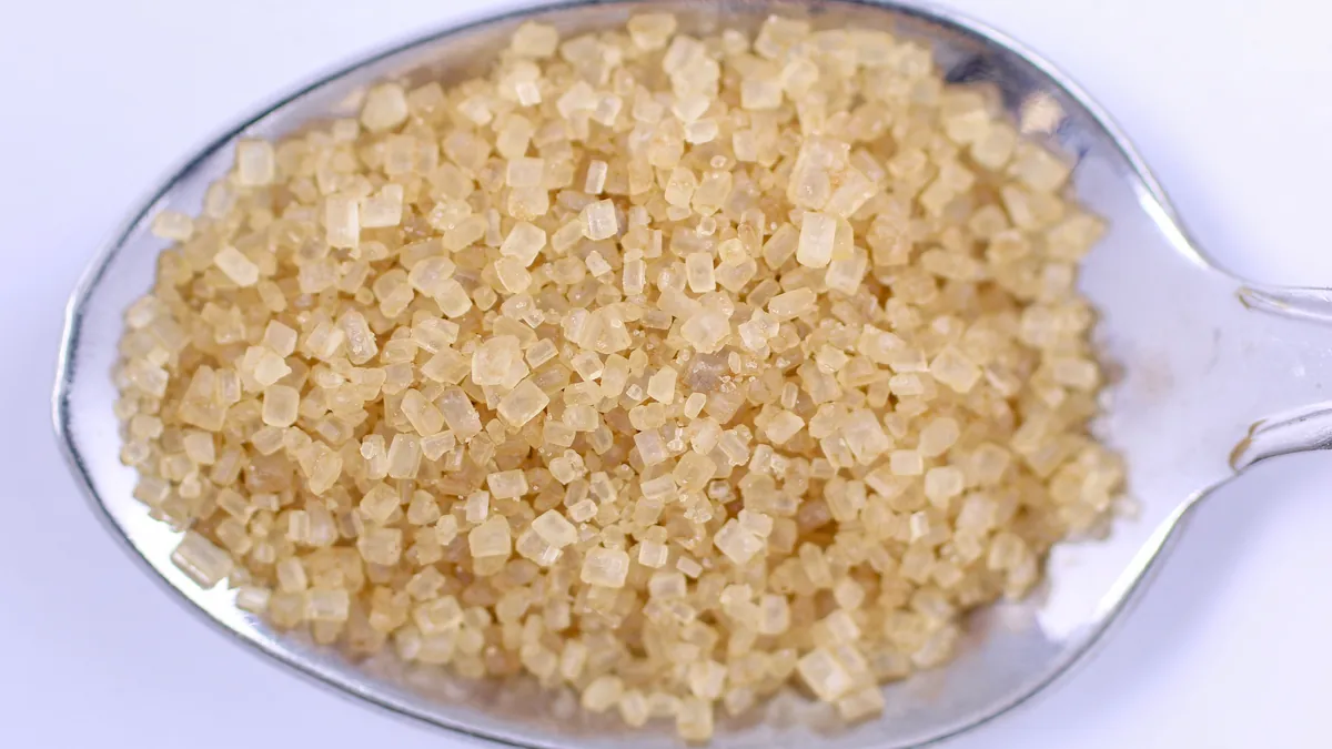
    In 1912, the French chemist Louis-Camille Maillard described what happens when sugars react with proteins: they bond together and form new, brown, rigid compounds. In cooking, that reaction creates flavor and color. In the body, the same reaction runs on your own tissues whenever glucose is abundant. Glucose molecules latch onto proteins — including the structural and functional proteins of nerves — and, through a series of steps, form stable end-products.

    These are called Advanced Glycation End-products, or AGEs — an apt acronym, because the process is a form of accelerated aging of the tissue. Once formed, AGEs are hard to remove, and they accumulate over years of elevated blood sugar.

    Three ways AGEs injure nerves

    Glycation harms nerves through several converging mechanisms.

    1. Structural cross-linking. AGEs bind proteins to one another, cross-linking them into stiff, dysfunctional complexes. In a nerve, this degrades the delicate architecture required to conduct signals and to maintain the insulating myelin sheath. Cross-linked proteins in the walls of the tiny blood vessels feeding the nerve also stiffen those vessels, choking the nerve’s blood supply.

    2. Inflammatory ignition through RAGE. AGEs are not just inert debris. They bind to a specific receptor called RAGE (the receptor for advanced glycation end-products) on the surface of cells. Activating RAGE switches on inflammatory signaling cascades, flooding the tissue with inflammatory mediators and oxidative stress. The nerve is effectively set on a low, chronic inflammatory fire.

    3. Oxidative stress and the polyol pathway. When glucose is abundant, some of it is shunted through the polyol pathway, where the enzyme aldose reductase converts glucose to sorbitol. This process consumes NADPH — the same molecule the cell needs to regenerate glutathione, its master antioxidant. The nerve is thus hit twice: sorbitol accumulates and draws water into the cell, while the antioxidant defense is depleted just as oxidative stress is rising.

    The unifying mechanism: mitochondrial overload

    How do these pathways connect? The landmark work of Dr. Michael Brownlee, published in Nature in 2001, proposed a unifying explanation: excess glucose overloads the mitochondria — the cell’s power plants — causing them to overproduce a damaging molecule called superoxide. That single upstream event, Brownlee argued, switches on the glycation, polyol, and inflammatory pathways together. In other words, mitochondrial overload is the common root, and AGEs, sorbitol accumulation, and RAGE-driven inflammation are its branches. This is why effective treatment has to consider mitochondrial health, not just blood sugar numbers in isolation.

    Why the feet burn first

    Nerves signal by maintaining a precise electrical and chemical environment along their length. As glycation stiffens their structure, inflammation irritates them, and oxidative stress and poor blood flow starve them of energy, the fibers begin to misfire — generating the burning, tingling, and electric sensations of neuropathy — and then to die back. Because the process is length-dependent, the longest nerves, which reach the feet, are affected first. That is the biochemical reason diabetic neuropathy characteristically begins in the toes and moves upward.

    Beyond the feet: the brain connection

    The reach of glucose-driven damage does not stop at the peripheral nerves. Research by Dr. Suzanne de la Monte at Brown University introduced the concept of Alzheimer’s disease as, in part, a metabolic disorder of brain insulin signaling — sometimes called “type 3 diabetes.” The same terrain of glycation, inflammation, and insulin resistance that injures peripheral nerves also appears to affect the brain, which is one reason chronic high blood sugar is associated with cognitive symptoms as well as neuropathy.

    Why a single glucose test isn’t enough

    A one-time fasting glucose is a snapshot; glycation is a movie. Because AGE formation depends on cumulative sugar exposure over time, better windows into the process include HbA1c (which reflects average glucose over roughly three months and is itself a glycated protein), a glucose tolerance test (which can reveal impaired glucose handling that fasting numbers miss), and markers of the downstream damage. Catching dysregulation at the prediabetes stage — before a formal diabetes diagnosis — matters, because nerve damage can begin there.

    What this means for treatment

    The biochemistry points directly at the strategy: reduce the ongoing glucose exposure driving glycation, support the antioxidant defenses (glutathione and its cofactors) that the polyol pathway depletes, and restore mitochondrial function so the upstream overload eases. This is the rationale behind a terrain-focused approach that pairs glycemic control with targeted metabolic and mitochondrial support, rather than relying only on drugs that mask the resulting pain.

    Frequently asked questions

    Can nerve damage from high blood sugar be undone?

    Some can, especially when caught early and when the underlying glucose exposure and oxidative stress are corrected. Established damage may only partly recover, so the priority becomes halting progression. Individual results vary.

    My fasting glucose is normal — am I in the clear?

    Not necessarily. HbA1c and a glucose tolerance test reveal patterns a single fasting number misses, and nerve damage can begin at the prediabetes stage.

    What are AGEs, in one sentence?

    Advanced glycation end-products are stiff, damaging compounds formed when sugar bonds to your proteins — the body’s internal version of caramelization.

    Does this affect anything besides my feet?

    Yes. The same metabolic terrain is linked to blood-vessel disease and to cognitive effects; research describes an insulin-resistance component of brain disease sometimes called “type 3 diabetes.”

    Key takeaways

    • The Maillard reaction bonds glucose to your proteins, forming stiff, damaging AGEs.
    • AGEs harm nerves by cross-linking structure, activating RAGE-driven inflammation, and driving oxidative stress via the polyol pathway.
    • Brownlee’s work identifies mitochondrial overload as the unifying upstream event.
    • Damage is length-dependent, so the feet are affected first.
    • HbA1c and glucose tolerance testing reveal the process better than a single fasting glucose.

    This article is educational and is not a substitute for evaluation, diagnosis, or treatment by a physician. Individual results vary. Do not start, stop, or change any medication without consulting your physician. Take the free Nerve Damage Score or call/text (314) 886-5902.

    References

    1. Maillard LC. Action des acides aminés sur les sucres; formation des mélanoïdines par voie méthodique. C R Acad Sci. 1912;154:66–68.
    2. Brownlee M. Biochemistry and molecular cell biology of diabetic complications. Nature. 2001;414:813–820.
    3. Vlassara H, Uribarri J. Advanced glycation end products (AGEs) and diabetes. Curr Diab Rep. 2014.
    4. de la Monte SM, Wands JR. Alzheimer’s disease is type 3 diabetes—evidence reviewed. J Diabetes Sci Technol. 2008;2(6):1101–1113.
    5. Oates PJ. Polyol pathway and diabetic peripheral neuropathy. Int Rev Neurobiol. 2002.

    Find out what is driving your nerve pain

    The free, five-question Nerve Damage Score takes about two minutes and tells you which terrain failure is most likely behind your symptoms.

    Get My Free Nerve Damage Score

    Or call or text (314) 886-5902.

  • Is Your Diet Destroying Your Nerves? The Gluten–Neuropathy Connection

    Is Your Diet Destroying Your Nerves? The Gluten–Neuropathy Connection

    Most people think of gluten as a digestive issue — a problem for the gut, felt as bloating or diarrhea. But some of the most important consequences of gluten sensitivity are neurological, and they can appear in people whose intestines seem perfectly fine. Gluten can injure the nervous system directly, producing peripheral neuropathy and balance problems even in the complete absence of celiac disease. This article explains the immune mechanism behind that, why standard celiac testing so often misses it, and what the evidence says about dietary elimination.

    Gluten sensitivity is not only celiac disease

    Celiac disease is an autoimmune reaction to gluten that damages the small intestine. But researchers — most prominently Dr. Marios Hadjivassiliou and colleagues in Sheffield, UK — have spent decades documenting a broader category of gluten-related disorders in which the primary target is not the gut but the nervous system. In many of these patients, intestinal biopsy is normal or near-normal, and yet the neurological damage is real and progressive.

    This matters enormously, because a patient with gluten neuropathy who is told “your celiac test is negative, so gluten isn’t your problem” may continue eating the very trigger that is dismantling their nerves.

    Molecular mimicry: how the immune system confuses nerves for gluten

    The central mechanism is molecular mimicry. When the immune system mounts a response to gluten, it produces antibodies. In susceptible people, some of those antibodies cross-react with the body’s own tissues because a molecular structure in nervous tissue resembles the gluten fragment closely enough to be mistaken for it.

    A key player is an enzyme family called transglutaminase. In celiac disease, the immune attack targets transglutaminase-2, concentrated in the gut. In gluten-related neurological disease, antibodies against transglutaminase-6 (TG6) — an isoform expressed in the nervous system — have been identified as a marker of the neural attack. Antibodies can also target the myelin that insulates nerves. The result is an immune assault on the peripheral nerves (neuropathy) and, in some patients, on the cerebellum, producing gluten ataxia — a loss of coordination and balance.

    What gluten neuropathy feels like

    Gluten neuropathy most often presents as a length-dependent sensory neuropathy: tingling, numbness, and burning that begin in the feet. Because it develops slowly and can occur without digestive symptoms, it is frequently mislabeled as idiopathic. When the cerebellum is involved, patients may notice unsteadiness, a wide-based gait, or clumsiness that they attribute to aging. The combination of an unexplained sensory neuropathy and subtle balance problems should raise the question of gluten sensitivity.

    Why the celiac test misses it

    Standard celiac screening looks for the intestinal form of the disease — typically tissue transglutaminase-2 (tTG) antibodies and evidence of gut damage. It is not designed to detect the neurological form. A patient can have a completely negative celiac panel and still be producing the TG6 and anti-myelin antibodies driving nerve injury. This is why a negative celiac test does not rule out gluten as a cause of neuropathy — a point explored further in the companion article on negative celiac testing.

    Genetic testing (for the HLA-DQ2/DQ8 haplotypes that predispose to gluten sensitivity) and expanded antibody panels give a more complete picture than a routine celiac screen alone.

    The role of strict elimination

    Because the damage is driven by an ongoing immune response to gluten, the logical — and evidence-supported — intervention is strict, sustained gluten elimination. Hadjivassiliou’s group has reported that patients who adhere rigorously to a gluten-free diet can see stabilization and, in some cases, improvement of their neurological symptoms, while partial or inconsistent avoidance is often insufficient. Nerve tissue recovers slowly, so improvement, when it comes, unfolds over months, and the earlier the trigger is removed, the more function there is to preserve.

    “Strict” is the operative word. Unlike a lifestyle preference, therapeutic gluten elimination for neurological disease means eliminating cross-contamination and hidden sources — not simply cutting back on bread. This is best done with dietary guidance and monitoring.

    Where this fits in a root-cause evaluation

    Gluten is one driver among several that can masquerade as idiopathic neuropathy. In a thorough workup it is evaluated alongside metabolic, toxic, and mechanical contributors, because more than one can be present at once. The value of identifying a gluten contribution is that it is, in principle, entirely removable — the trigger is on the plate.

    Frequently asked questions

    Can I have gluten neuropathy if my celiac test is negative?

    Yes. Standard celiac tests detect the intestinal form of gluten disease, not the neurological form. TG6 and anti-myelin antibodies can be present with a negative celiac panel.

    Do I need digestive symptoms to have gluten neuropathy?

    No. Many patients with gluten-related nerve damage have no significant gut symptoms at all.

    Will going gluten-free fix my neuropathy?

    It can help stabilize and sometimes improve symptoms when done strictly and early, but nerve recovery is slow and varies by individual. It is most effective as part of a physician-guided plan.

    How strict do I need to be?

    For neurological gluten disease, strict elimination — including hidden and cross-contaminating sources — appears necessary; occasional avoidance is generally not enough.

    Key takeaways

    • Gluten can injure nerves and the cerebellum directly, independent of celiac disease.
    • The mechanism is immune molecular mimicry, with transglutaminase-6 antibodies marking the neural attack.
    • Standard celiac tests do not detect the neurological form; a negative result does not clear gluten.
    • Strict, sustained gluten elimination is the evidence-supported intervention, with slow, partial recovery possible.
    • Gluten is evaluated alongside other neuropathy drivers, and is uniquely removable.

    This article is educational and is not a substitute for evaluation, diagnosis, or treatment by a physician. Individual results vary. Do not make major dietary or medication changes without consulting your physician. Take the free Nerve Damage Score or call/text (314) 886-5902.

    References

    1. Hadjivassiliou M, Grünewald RA, Davies-Jones GAB. Gluten sensitivity as a neurological illness. J Neurol Neurosurg Psychiatry. 2002;72:560–563.
    2. Hadjivassiliou M, et al. Transglutaminase-6 antibodies in the diagnosis of gluten ataxia. Neurology. 2013;80:1740–1745.
    3. Hadjivassiliou M, et al. Gluten neuropathy. Muscle Nerve / Brain (peripheral neuropathy series).
    4. Hadjivassiliou M, et al. Neurologic deficits in patients with gluten sensitivity — role of strict gluten-free diet.

    Note: match each reference to the specific paper at publication; the Sheffield group has an extensive body of work in this area.

    Find out what is driving your nerve pain

    The free, five-question Nerve Damage Score takes about two minutes and tells you which terrain failure is most likely behind your symptoms.

    Get My Free Nerve Damage Score

    Or call or text (314) 886-5902.

  • The Neuropathy Breakthrough Mainstream Medicine Overlooked: The High-Concentration Capsaicin Protocol

    The Neuropathy Breakthrough Mainstream Medicine Overlooked: The High-Concentration Capsaicin Protocol

    There is a treatment for neuropathic foot pain that is FDA-approved, non-opioid, non-systemic, and applied in a doctor’s office in under an hour — and yet many patients living with burning feet have never been offered it. It is the high-concentration 8% capsaicin patch. This article explains how it works, what the clinical evidence genuinely shows, why it often takes more than one treatment to see the full benefit, and how it fits into a repair-focused protocol. It also draws a careful line between what is proven and what is still being studied — because that distinction is exactly what “mainstream medicine missed” conversations tend to blur.

    A note on numbers, up front

    You may have seen a very high success figure attached to this protocol. In the interest of honesty: the controlled clinical trials of the 8% capsaicin patch show modest but statistically significant pain relief, not near-universal cure. Any higher real-world success rate reported by an individual clinic reflects that clinic’s own patient population, protocol, and outcome definition — it is a practice observation, not a figure from the randomized trials, and it should be understood that way. What follows sticks to the published science, then explains where clinical experience extends beyond it.

    The receptor that makes chili peppers hot

    Capsaicin is the compound that gives chili peppers their heat. It acts on a specific sensor on pain-sensing nerve endings called the TRPV1 receptor (transient receptor potential vanilloid 1). TRPV1 is the same receptor that responds to noxious heat — which is why capsaicin literally feels hot.

    Low-dose capsaicin creams (0.025–0.075%) available over the counter provide mild, temporary relief and must be applied several times a day. The prescription 8% patch is a different order of magnitude — roughly a hundredfold more concentrated — and is designed to do something the creams cannot: temporarily defunctionalize the overactive pain fibers.

    Defunctionalization: turning down an alarm that won’t stop

    In chronic neuropathy, the small C-fibers in the skin become pathologically hyperexcitable. They fire pain signals continuously, even without a real stimulus — the alarm is stuck on. A single, controlled 30-minute application of the 8% patch delivers enough capsaicin to overstimulate those TRPV1-bearing endings and then reversibly quiet them. The nerve endings retract and stop transmitting their runaway pain signals; over the following weeks and months they gradually recover. Crucially, controlled studies found this happens without degrading normal sensation — patients retained their ability to feel sharp, warm, cold, and vibration stimuli.

    Because the treatment is topical and non-systemic, it sidesteps the sedation, dizziness, and cognitive fog that limit oral neuropathic-pain drugs. The main side effect is temporary burning and redness at the application site, which is why the patch is applied in a clinical setting, often with skin pre-treatment for comfort.

    What the evidence shows

    The 8% patch first earned FDA approval in 2009 for postherpetic neuralgia (the lingering nerve pain after shingles). In 2020, on the strength of the pivotal STEP trial led by Dr. David Simpson, the FDA extended approval to painful diabetic peripheral neuropathy of the feet. In STEP, a single 30-minute treatment produced a statistically significant reduction in average daily pain compared with placebo. A separate 52-week study (PACE) confirmed that repeated treatments were well tolerated over a full year, with no worsening of sensory function.

    This is the honest headline: the patch reliably and safely reduces pain for a meaningful share of patients, and it can be repeated approximately every three months.

    Why persistence matters

    One reason patients — and sometimes physicians — give up too soon is that they expect a single patch to do everything. The data and clinical experience both suggest the benefit often builds across successive cycles. Each treatment quiets the overfiring fibers again and extends the window of reduced pain. For a chronic condition that took years to develop, expecting resolution from one application is unrealistic; the protocol is designed around repeated, spaced treatments.

    Can it help nerves regrow?

    Here is where the science is genuinely exciting but not yet settled. Because capsaicin causes nerve endings to retract and then regenerate, researchers have asked whether repeated treatment might not just relieve pain but actually encourage healthier re-innervation of the skin — in other words, whether it could be disease-modifying. A randomized trial in the United Kingdom is formally testing exactly this question in diabetic neuropathy.

    Until that kind of study reports, the responsible position is: the 8% patch is proven for pain relief, and its potential to modify the underlying nerve damage is a promising hypothesis under active investigation. Regenerve’s approach uses the pain-relief window the patch creates as the foundation for the repair-focused steps — orthobiologics and mitochondrial and metabolic support — that aim at the nerve itself. Those regenerative components, as covered in the protocol article, are themselves emerging and individualized rather than guaranteed.

    Who is a candidate

    The patch is used for localized neuropathic pain, classically in the feet for diabetic neuropathy and in the affected area for postherpetic neuralgia. Suitability depends on a physician’s evaluation of the pain distribution, skin integrity, and overall picture. As with any treatment, it works best as part of a plan that also addresses the root drivers of the neuropathy rather than as a standalone fix.

    Frequently asked questions

    Does the capsaicin patch cure neuropathy?

    No. It reduces neuropathic pain — often meaningfully and safely — and can be repeated. It is not a cure, and its ability to reverse nerve damage is still under investigation.

    How is it different from capsaicin cream?

    The 8% patch is prescription-strength, applied once in a clinical setting for about 30 minutes, and roughly 100 times more concentrated than over-the-counter creams.

    Does it hurt?

    There is temporary burning and redness at the site during and shortly after application. It is applied under supervision, often with steps taken to improve comfort, and does not damage normal sensation.

    How often can it be repeated?

    Approximately every three months, and benefit often accumulates over successive treatments.

    What about that very high success rate I saw?

    Controlled trials show modest, significant relief. Higher figures reported by a clinic reflect its own real-world experience and outcome definitions, not the randomized-trial data, and should be interpreted with that context.

    Key takeaways

    • The 8% capsaicin patch is FDA-approved for painful diabetic neuropathy of the feet (2020) and postherpetic neuralgia (2009).
    • It works by reversibly quieting overfiring TRPV1 pain fibers, without degrading normal sensation, and without systemic side effects.
    • Trial evidence supports real but modest pain relief; benefit often builds across repeated treatments.
    • Whether it can help nerves regenerate is a legitimate, still-unproven hypothesis under study.
    • It works best inside a plan that also treats the neuropathy’s root causes.

    This article is educational and is not a substitute for evaluation, diagnosis, or treatment by a physician. Individual results vary. Do not start, stop, or change any treatment without consulting your physician. Take the free Nerve Damage Score or call/text (314) 886-5902.

    References

    1. Simpson DM, Robinson-Papp J, Van J, et al. Capsaicin 8% patch in painful diabetic peripheral neuropathy: a randomized, double-blind, placebo-controlled study (STEP). J Pain. 2017;18(1):42–53.
    2. U.S. FDA. Qutenza (capsaicin) 8% topical system — approval for painful diabetic peripheral neuropathy of the feet. 2020; original PHN approval 2009.
    3. Vinik AI, et al. Capsaicin 8% patch repeat treatment plus standard of care in painful diabetic peripheral neuropathy: 52-week open-label safety study (PACE). BMC Neurol. 2016;16:251.
    4. Anand P, Bley K. Topical capsaicin for pain management: mechanisms of action of the 8% capsaicin patch. Br J Anaesth. 2011;107(4):490–502.
    5. Abrams RMC, et al. A critical review of the capsaicin 8% patch for diabetic peripheral neuropathy of the feet. Expert Rev Neurother. 2021.

    Find out what is driving your nerve pain

    The free, five-question Nerve Damage Score takes about two minutes and tells you which terrain failure is most likely behind your symptoms.

    Get My Free Nerve Damage Score

    Or call or text (314) 886-5902.

  • Peripheral Neuropathy: The 11 Hidden Drivers Your Doctor Missed

    Peripheral Neuropathy: The 11 Hidden Drivers Your Doctor Missed

    When a workup comes back inconclusive, many patients are told their neuropathy is “idiopathic” — medical language for cause unknown. But in a large share of these cases, the cause isn’t truly unknown; it simply hasn’t been looked for thoroughly enough. Idiopathic is too often a clinical surrender rather than a diagnosis. This article lays out the systematic investigation Dr. Padda uses to hunt down the real driver of nerve damage, organized into three domains — metabolic, toxic, and mechanical — because a nerve can die from many directions, and finding which one is the difference between managing symptoms forever and actually changing course.

    “Idiopathic” is often incomplete, not unsolvable

    Studies of unexplained peripheral neuropathy consistently show that when patients undergo a structured, expanded evaluation, an identifiable cause emerges in a substantial proportion of cases previously labeled idiopathic. The American Academy of Neurology’s evaluation guidance for distal symmetric polyneuropathy emphasizes a tiered laboratory workup precisely because targeted testing changes management. The point is not that every case has an easy answer — some remain genuinely unexplained — but that the label should be earned only after a real search.

    The three-domain framework below is a way to organize that search so nothing obvious gets skipped.

    Domain one: metabolic drivers

    Metabolic problems are the most common and most treatable causes of nerve damage. They share a final common pathway — energy failure and chemical injury inside the nerve.

    1. Blood sugar and glycation. Diabetes is the leading cause of neuropathy worldwide, but the damage begins earlier than most people realize. Research from the University of Utah (Drs. Smith and Singleton) helped establish that even prediabetes — impaired glucose tolerance that never reaches the diabetes threshold — is associated with small-fiber neuropathy. Chronically elevated glucose bonds to nerve proteins to form advanced glycation end-products through the Maillard reaction, first described by Louis-Camille Maillard in 1912. A standard fasting glucose can miss this entirely; markers like HbA1c and a glucose tolerance test reveal far more. (Explored in depth in the glycation and blood-sugar articles.)

    2. Intracellular nutritional deficiency. Nerves are cofactor-hungry. Deficiencies of B1 (thiamine), B6, B12, folate, magnesium, and omega-3 fatty acids each impair nerve function and mitochondrial energy production. Crucially, blood levels can look “normal” while the tissue is starved — which is why functional testing matters (see driver 3).

    3. Functional B12 deficiency. A serum B12 in the normal range does not rule out a cellular deficiency. Functional markers — methylmalonic acid (MMA) and homocysteine — rise when B12 is functionally inadequate at the tissue level, catching deficiencies a standard B12 test misses. Untreated, B12 deficiency causes a characteristic neuropathy (and can damage the spinal cord), yet it is eminently correctable. (Covered fully in the B12 article.)

    Domain two: toxic drivers

    If metabolic drivers are about deprivation, toxic drivers are about poisoning — substances that damage nerves directly or by depleting the body’s defenses.

    4. Bioaccumulated heavy metals. Lead, arsenic, mercury, and thallium disrupt essential enzymes and deplete glutathione, the body’s master antioxidant. Exposure is often occupational or environmental and accumulates silently over years. Testing should be guided by a genuine exposure history — and, importantly, patients should be steered away from unproven or aggressive “detox” schemes, which can do more harm than good. (See the heavy-metals article.)

    5. Mold and mycotoxins. Toxins produced by mold in water-damaged buildings can impair mitochondrial function and drive neuroinflammation, sometimes presenting as neuropathy paired with profound fatigue. This is a genuinely debated clinical area, and it deserves careful, evidence-aware evaluation rather than either dismissal or overdiagnosis. (See the mycotoxin article.)

    6. Neurotoxic medications. Some of the most commonly prescribed drugs can quietly undermine nerve health. Statins can deplete CoQ10; metformin — one of the most-prescribed diabetes drugs — can cause B12 deficiency over time; certain chemotherapy agents and antibiotics are directly neurotoxic. The answer is rarely to stop a needed medication, but to monitor and replete the nutrients they affect. (See the statins-and-metformin article.)

    7. Alcohol. Alcohol is a double hit: acetaldehyde and alcohol itself are directly toxic to nerves, and heavy use depletes thiamine and other B vitamins. Recovery depends on reducing exposure and repleting nutrients — but abrupt cessation in someone alcohol-dependent carries its own medical risks and should be handled with clinical guidance. (See the alcohol article.)

    Domain three: mechanical and immune drivers

    The final domain covers physical and immune-mediated injury — nerves crushed, choked, or caught in the crossfire of the body’s own defenses.

    8. Autoimmune disease. Conditions like lupus, rheumatoid arthritis, and Sjögren’s damage nerves through vasculitis (inflammation of the small vessels feeding the nerve) and direct antibody attack. Here the neuropathy is collateral damage from a systemic process, so treatment requires controlling the underlying disease and addressing the nerve. (See the autoimmune article.)

    9. Gluten sensitivity. Even without celiac disease, gluten can trigger a neurological immune response — antibodies against transglutaminase-6 have been linked to neuropathy and cerebellar ataxia in work led by Dr. Marios Hadjivassiliou. A negative celiac test does not rule this out. (See the two gluten articles.)

    10. Chronic stealth infections. Certain infections hide in nervous tissue. The varicella-zoster (shingles) virus resides in the dorsal root ganglia and can reactivate to cause postherpetic neuralgia; Lyme and other pathogens can also produce neuropathy. (See the shingles article.)

    11. Mechanical compression. Sometimes it isn’t a metabolic disease at all — it’s a pinched nerve. Carpal tunnel syndrome, radiculopathy (sciatica), and other entrapments injure nerves through ischemia and focal demyelination. The concept of double crush syndrome, described by Upton and McComas in 1973, explains why a metabolically stressed nerve is more vulnerable to a second, mechanical injury — meaning compression and metabolic disease often compound each other. (See the compression article.)

    Why one patient often has several drivers at once

    These categories are not mutually exclusive. A person with prediabetes, a statin prescription, and a compressed nerve at the wrist may have three simultaneous drivers, each amplifying the others. This is the practical reason a single-cause mindset fails: the workup has to be broad enough to catch combinations, and the treatment plan has to address all the active contributors, not just the most obvious one.

    What a thorough workup looks like

    A genuine root-cause investigation typically includes a detailed history (occupation, exposures, diet, alcohol, medications, family history), an expanded metabolic panel (glucose tolerance and HbA1c, not just fasting glucose), functional nutrient testing (MMA and homocysteine, thiamine, magnesium), targeted autoimmune and infectious testing when the history points that way, and — where relevant — nerve conduction studies or skin biopsy to characterize the fiber types involved. The aim is to convert “idiopathic” into a named, addressable cause whenever the evidence allows.

    Frequently asked questions

    My tests were normal — does that mean there’s no cause?

    Not necessarily. Standard panels can miss functional deficiencies (like tissue-level B12), early glucose dysregulation, and toxic exposures. A broader, targeted workup often uncovers a driver that routine testing skips.

    That is the subject of Mystery Neuropathy and Exhaustion? The Mold and Mycotoxin Question.

    Can more than one thing be causing my neuropathy?

    Yes, and it’s common. Multiple drivers frequently coexist and compound one another, which is why a complete evaluation matters.

    This is set out in The Sugar That Caramelizes Your Nerves: How Glucose Damages Your Nervous System.

    Is it too late if I’ve had neuropathy for years?

    Identifying and removing an active driver can slow or halt progression at any stage, and some function may recover. Earlier is better, but a workup is worthwhile regardless of how long symptoms have been present.

    There is more on this in Your “Normal” B12 May Be Lying: The Hidden Deficiency Driving Your Neuropathy.

    Should I try a detox for heavy metals?

    Only under medical guidance and only if testing and history justify it. Unproven chelation and “detox” protocols carry real risks and should not be self-administered.

    Mystery Neuropathy and Exhaustion? The Mold and Mycotoxin Question explains what that looks like.

    Key takeaways

    • “Idiopathic” often means the search was incomplete, not that no cause exists.
    • Nerve damage arises from metabolic, toxic, and mechanical drivers — often several at once.
    • Standard labs miss a lot: functional B12 markers, glucose tolerance, and exposure-guided testing reveal more.
    • Double crush syndrome shows how metabolic and mechanical injuries compound each other.
    • The goal of the workup is to convert an unexplained neuropathy into a named, treatable one.

    This article is educational and is not a substitute for evaluation, diagnosis, or treatment by a physician. Individual results vary. Do not start, stop, or change any medication or treatment without consulting your physician. Take the free Nerve Damage Score or call/text (314) 886-5902 to begin a root-cause evaluation.

    References

    1. England JD, Gronseth GS, Franklin G, et al. Distal symmetric polyneuropathy: a definition for clinical research (AAN/AANEM/AAPM&R). Neurology. 2005; and the AAN evaluation guidance, Neurology. 2009.
    2. Smith AG, Singleton JR. Impaired glucose tolerance and neuropathy. Neurologist / Diabetes Care (University of Utah body of work).
    3. Maillard LC. Action des acides aminés sur les sucres. C R Acad Sci. 1912.
    4. Brownlee M. Biochemistry and molecular cell biology of diabetic complications. Nature. 2001;414:813–820.
    5. Hadjivassiliou M, et al. Transglutaminase-6 antibodies and gluten-related neurological dysfunction. Neurology / Ann Neurol.
    6. Upton ARM, McComas AJ. The double crush in nerve-entrapment syndromes. Lancet. 1973;2(7825):359–362.

    Note: match each reference to a specific, current source at publication; several point to bodies of work rather than a single paper.

    Find out what is driving your nerve pain

    The free, five-question Nerve Damage Score takes about two minutes and tells you which terrain failure is most likely behind your symptoms.

    Get My Free Nerve Damage Score

    Or call or text (314) 886-5902.