
Category: Diabetic & Metabolic Drivers

  • Neuropathy Before a Diabetes Diagnosis: Prediabetes and Nerve Damage Explained (2026)

    Neuropathy Before a Diabetes Diagnosis: Prediabetes and Nerve Damage Explained (2026)

    Nerve symptoms can begin before anyone tells you that you have diabetes, and there are two separate reasons for that. The first is that a lot of diabetes is simply not yet identified: the CDC estimates that 27.6% of US adults aged 18 and older who have diabetes are undiagnosed, which is about 11.0 million people [1]. The second is that nerve injury is reported in people whose glucose handling is impaired but has not crossed the diabetes threshold [2].

    Key takeaways

    • An estimated 27.6% of US adults with diabetes do not know they have it, roughly 11.0 million people, based on 2023 data [1].
    • An estimated 115.2 million US adults aged 18 and older have prediabetes [1].
    • Peripheral neuropathy has been reported more frequently in people with impaired glucose tolerance than in people with normal glucose tolerance [2].
    • About 20% of people with type 2 diabetes already have diabetic peripheral neuropathy at the time the diabetes is diagnosed [2].
    • Neuropathy is a category, not a diagnosis. Glucose is one driver among several, and more than one is often active at once.
    • Reduced protective sensation is a safety problem before it is a comfort problem. Daily foot checks start now, not after testing.

    Why the timeline is confusing

    People reasonably assume the sequence runs diagnosis first, complications later. In practice the metabolic changes precede the label, sometimes by years, and the label arrives when someone happens to run the right test.

    That is why “my sugar was normal” is a weaker reassurance than it sounds. It describes one measurement on one day, and the CDC estimate of 11.0 million US adults with undiagnosed diabetes shows how often the measurement has simply not been made [1].

    What the prediabetes research actually reports

    A 2017 review in the Journal of Diabetes Investigation examined peripheral neuropathy in prediabetes and the metabolic syndrome. It reports that neuropathy has been found more often in people with impaired glucose tolerance than in people with normal glucose tolerance, drawing on population studies that examined all three groups [2].

    The same review notes that roughly 20% of people with type 2 diabetes already have diabetic peripheral neuropathy at the point the diabetes is diagnosed [2]. Nerve involvement is not a late complication in those cases; it is present at the start.

    What this does not establish is a threshold below which nerves are safe. It establishes that glucose handling short of diabetes is worth taking seriously when someone has foot symptoms.

    What happens around the nerve

    Peripheral nerves are supplied by very small blood vessels and depend on steady oxygen delivery and stable metabolic conditions. Both of those can be disturbed while a person is still described as not diabetic.

    Glycation

    Glucose reacts with proteins over time, and the products of that reaction accumulate in tissue. This is the chemistry behind how sugar damages nerves, and it does not switch on at a diagnostic cutoff.

    Perfusion and metabolic strain

    Nerve tissue has a high energy demand relative to its blood supply. When delivery and demand stop matching, the longest fibers, the ones reaching the toes, are the first to show it, which is why symptoms usually start at the feet.

    Symptoms worth taking to a clinician

    The pattern matters more than the intensity. What people describe most often is burning at night, pins and needles, numbness that spreads slowly upward from the toes, or a sense that the foot is less responsive than it used to be.

    Numbness is a safety issue first

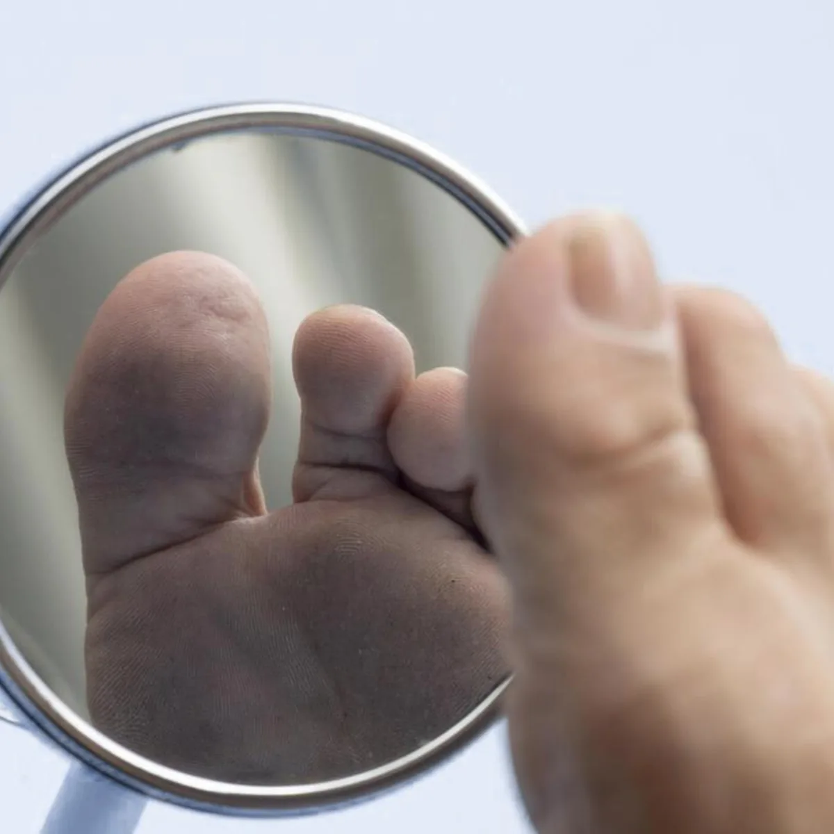
    Loss of protective sensation means a blister, a seam, a pebble or a burn can go unnoticed until the skin has broken down. Check both feet daily, including between the toes and across the soles, and use a mirror or a second pair of eyes if you cannot see them easily.

    Any open area, or a foot that is hot and swollen, needs evaluation rather than observation.

    Hand holding a round mirror to inspect the sole of a foot, the daily self-check recommended when protective sensation is reduced by peripheral neuropathy

    Glucose is one driver, not the whole list

    Even when glucose handling is clearly involved, it is rarely the only thing acting on the nerve. A work-up that stops at the metabolic answer will miss the contributors that are easiest to correct.

    • Nutritional, including B12 status and malabsorption after gastrointestinal surgery.
    • Autoimmune and inflammatory, including gluten-related and connective tissue processes.
    • Toxic, including alcohol, heavy metal exposure and medication effects such as chemotherapy.
    • Structural, including nerve entrapment and spine-related contributions.

    How the evaluation is sequenced at Regenerve

    The order is deliberate: establish the symptom pattern, then measure, then decide. Electrodiagnostic testing (EMG/NCS) is performed on site and characterizes conduction in the larger nerve fibers.

    When the story is burning and temperature change rather than numbness and weakness, the small fibers are the likely target, and conduction studies are not the right instrument for that. We use in-office small-fiber and vascular assessment in those cases, alongside metabolic and nutritional evaluation.

    What care addresses, and what it does not promise

    Regenerve offers class 4 photobiomodulation, class 3B cold laser, whole-body infrared and metabolic and nutritional care as components of a plan built around the identified driver. Orthobiologic injections such as PRP and BMAC are offered in selected situations.

    We do not promise nerve regeneration, reversal, and we do not publish success percentages for our protocols.

    Frequently asked questions

    Can nerve damage start before diabetes is diagnosed?

    It can. A review of peripheral neuropathy in prediabetes and the metabolic syndrome reports that neuropathy has been found more often in people with impaired glucose tolerance than in people with normal glucose tolerance, and that roughly 20% of people with type 2 diabetes already have diabetic peripheral neuropathy when the diabetes is first identified. See how blood sugar damages nerves.

    My A1c is normal. Does that rule out a metabolic cause?

    Not by itself. A single value in the normal range describes that moment rather than the years behind it, and glucose is only one of several metabolic inputs a nerve depends on. The more useful question is what else is on the list. See the hidden drivers of peripheral neuropathy.

    What else causes neuropathy when blood sugar looks fine?

    Nutritional, autoimmune, toxic and structural causes all produce foot symptoms that resemble the metabolic pattern, and more than one is often present at the same time. B12 status is one of the first to check because it is common, testable and correctable. See functional B12 deficiency and neuropathy.

    Should I be checked for celiac disease or gluten sensitivity?

    It belongs in the work-up when the pattern does not fit a length-dependent metabolic neuropathy, or when digestive symptoms sit alongside the nerve symptoms. Standard screening does not close the question in every case. See the gluten and neuropathy connection.

    My feet are numb. What should I be doing at home right now?

    Check both feet daily, including between the toes and the soles, because reduced protective sensation means an injury can go unnoticed until it is infected. Use a mirror or ask someone to look, and get any open area or hot, swollen foot evaluated rather than watched. See small fiber neuropathy symptoms and testing.

    Start with the Nerve Damage Score

    The entry point at Regenerve is the free five-question Nerve Damage Score. It takes a few minutes, and it tells us which driver to look at first so your visit starts with a plan instead of a guess.

    Take the free Nerve Damage Score assessment, then bring the result to a physician-led visit at Regenerve, 4477 Woodson Rd #104, St. Louis, MO 63134, minutes from St. Louis Lambert International Airport. We see patients from the St. Louis region, Missouri and Illinois.

    Sources

    1. Centers for Disease Control and Prevention. National Diabetes Statistics Report, updated January 21, 2026 (2023 data). https://www.cdc.gov/diabetes/php/data-research/index.html — referenced for the estimate that 27.6% of US adults aged 18 or older with diabetes are undiagnosed, representing 11.0 million people, and for the estimate of 115.2 million US adults aged 18 or older with prediabetes.
    2. Stino AM, Smith AG. “Peripheral neuropathy in prediabetes and the metabolic syndrome.” Journal of Diabetes Investigation, 2017. https://pmc.ncbi.nlm.nih.gov/articles/PMC5583955/ — referenced for the higher reported frequency of neuropathy in people with impaired glucose tolerance compared with people with normal glucose tolerance, and for the finding that approximately 20% of people with type 2 diabetes have diabetic peripheral neuropathy at the time of diagnosis.
  • Early signs of diabetic peripheral neuropathy: burning feet at night, numbness, and what to do

    Early signs of diabetic peripheral neuropathy: burning feet at night, numbness, and what to do

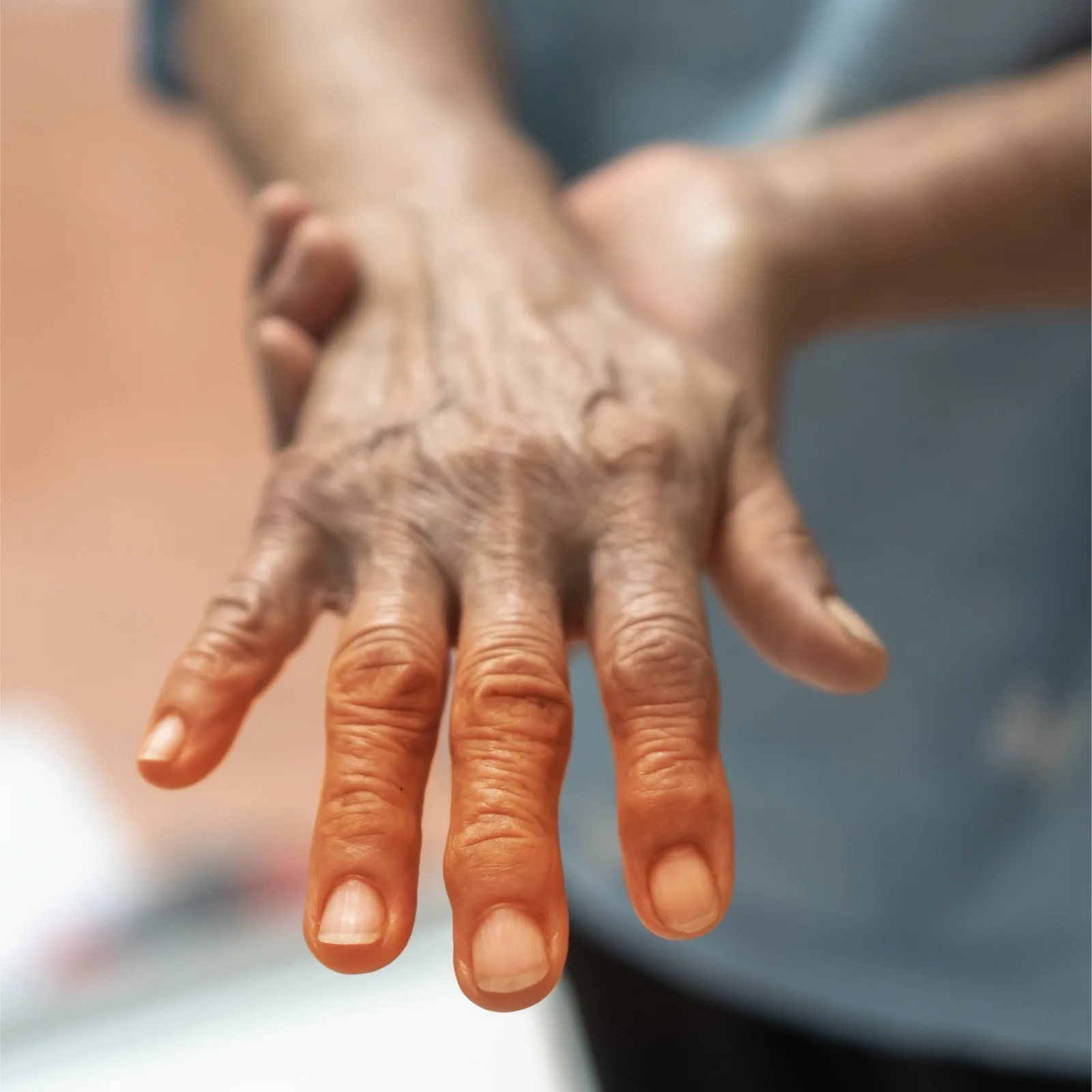
    The early signs are sensory and they start in the toes: burning at night, tingling, and numbness that spreads upward on both feet at once. Diabetic peripheral neuropathy occurs in up to 50% of patients with diabetes mellitus and commonly presents as distal symmetric polyneuropathy with a stocking-and-glove distribution, starting in the toes and moving proximally.1 Recognizing the pattern early matters because the same paper links it to loss of protective sensation and the cascade into foot ulcers, wounds and infections.1

    Older adult's outstretched hand with the fingertips shaded, showing the glove pattern of distal symmetric peripheral neuropathy

    What the early pattern looks like

    These changes rarely feel like an emergency at first. People describe them as being in the socks, on the skin, or in the floor underfoot.

    • Burning in the feet that is worse at night or after lying down
    • Tingling or pins-and-needles in the toes or forefoot
    • Numbness that starts small and spreads upward
    • Brief electric or stabbing pains
    • Unsteadiness on uneven ground or in the dark

    The symmetry is the diagnostic clue. A problem in one foot alone points somewhere else; the same change in both feet, worst at the toes, fits a length-dependent process.

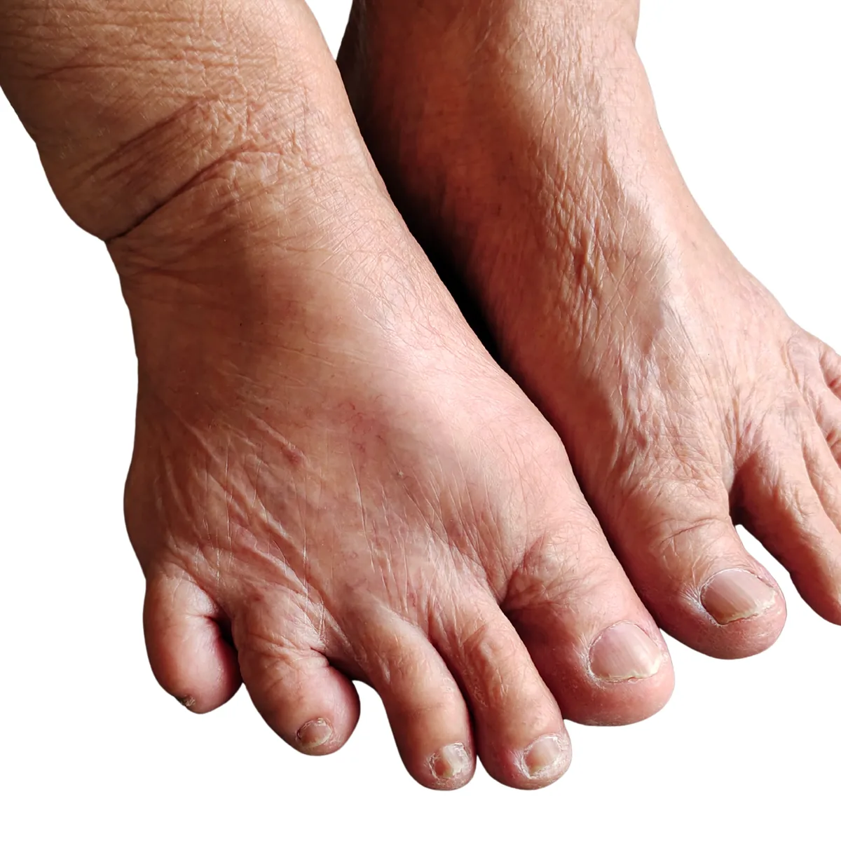
    Bare feet of an older adult showing dry skin, thickened toenails and bunion deformity, the appearance often seen alongside peripheral neuropathy of the feet

    Why numbness is the more dangerous symptom

    Pain gets attention. Numbness does not, which is exactly the problem: American Family Physician notes that diabetic peripheral neuropathy may result in a loss of protective sensation and cascade into foot ulcers, wounds, infections and tissue necrosis.1

    Once protective sensation is reduced, a blister or a stone in the shoe no longer announces itself. That is the point at which daily foot inspection stops being optional.

    Hand holding a round mirror to inspect the sole of a foot, the daily self-check recommended when protective sensation is reduced by peripheral neuropathy

    Why a normal A1C does not settle the question

    Fasting glucose and A1C describe circulating sugar at a point in time. They are not a direct measure of the metabolic state around the nerve, and a result inside the reference range does not by itself rule out an early neuropathic process.

    In our clinic we look at insulin resistance and the wider metabolic picture rather than relying on A1C alone. The reasoning is set out in blood sugar and nerve damage, and why normal labs miss it.

    Where chronic hyperglycemia is present, glycation is one of the mechanisms by which sugar damages nerve tissue over time. We explain it in how glycation damages nerves.

    Other drivers that can be present at the same time

    Neuropathy is a category, not a diagnosis, and a diabetes diagnosis does not exclude a second cause. American Family Physician specifically directs physicians to address underlying risk factors including vitamin B12 deficiency alongside glycemic control.1

    Nutritional, autoimmune, toxic and structural drivers all produce foot symptoms, and treating only the one you assumed is how a plan stalls. Identifying which driver is doing the most damage is the first job of the evaluation.

    What evaluation and care look like at Regenerve

    We perform electrodiagnostic testing (EMG/NCS) on site, and we sort between metabolic, nutritional, autoimmune, toxic and structural drivers before building a plan. Where balance is involved, VNG is used as a diagnostic test of inner-ear balance function, not as a treatment.

    Care options include physician-directed metabolic and nutritional care, class 4 photobiomodulation, class 3B cold laser and whole-body infrared. Orthobiologic injections (PRP and BMAC) are offered in selected situations. Qutenza (capsaicin 8% patch) is FDA-approved in adults only for neuropathic pain from postherpetic neuralgia and from diabetic peripheral neuropathy of the feet; it is applied in clinic by a clinician and repeated no more often than every three months.

    We do not promise nerve regeneration, reversal or cure, and we do not publish a success rate for our protocol. We are at 4477 Woodson Rd #104, St. Louis, MO 63134, and we see patients from the St. Louis region, Missouri and Illinois.

    What to do this week

    • Check both feet daily, soles included, using a mirror if you cannot see them.
    • Write down when the burning is worst and whether it is spreading.
    • Wear shoes that do not create pressure points, and check inside them before putting them on.
    • Ask for a work-up that looks past A1C, including B12 status.
    • Get evaluated rather than waiting for the pain to make the decision for you.

    Start with your Nerve Damage Score

    If you recognize the pattern, do not wait for it to spread. The Nerve Damage Score is a free five-question screen that helps identify which driver to test for first.

    Frequently asked questions

    What are the earliest signs of diabetic peripheral neuropathy?

    Sensory changes in the toes come first: burning at night, tingling, and numbness that spreads upward in a symmetric pattern. American Family Physician describes the presentation as distal symmetric polyneuropathy with a stocking-and-glove distribution, starting in the toes and moving proximally. See peripheral neuropathy of the feet symptoms.

    My nerve testing was normal but my feet burn at night. What now?

    Standard nerve conduction studies are weighted toward large myelinated fibers, so a small-fiber problem can sit beneath a normal result. Burning, temperature changes and altered sweating are the pattern worth raising. See small fiber neuropathy symptoms and testing.

    Can I have nerve symptoms from something other than blood sugar?

    Yes, and more than one driver can be present at the same time. Nutritional, autoimmune, toxic and structural causes all produce foot symptoms, which is why the work-up looks past the diabetes label. See why a normal B12 result can still miss neuropathy.

    Is numbness better than pain?

    No. Loss of protective sensation is what allows an injury to go unnoticed, and American Family Physician links it to the development of foot ulcers, wounds and infections. Numbness is a reason to start daily foot checks, not to relax. See exercises for peripheral neuropathy in the feet.

    Sources

    1. Bragg S, Marrison ST, Haley S. “Diabetic Peripheral Neuropathy: Prevention and Treatment.” American Family Physician. 2024;109(3):226. https://www.aafp.org/afp/2024/0300/diabetic-peripheral-neuropathy
    2. Hicks CW, Selvin E. “Epidemiology of Peripheral Neuropathy and Lower Extremity Disease in Diabetes.” Current Diabetes Reports. 2019;19(10):86. https://pmc.ncbi.nlm.nih.gov/articles/PMC6755905/
  • Diabetic Foot Ulcer Prevention When You Have Neuropathy

    Diabetic Foot Ulcer Prevention When You Have Neuropathy

    When neuropathy has taken your protective sensation, foot ulcer prevention stops being a hygiene habit and becomes a substitution: you replace the warning your nerves used to give with a daily visual check and deliberate control of pressure. Diabetic peripheral neuropathy occurs in up to 50% of patients with diabetes mellitus [1][4], and in that group an injury can develop well before anything hurts.

    That is the whole logic of prevention here. Look, because you can no longer feel; and control the pressure and friction that create the injury in the first place.

    Why neuropathy changes the arithmetic

    Metabolic neuropathy is length-dependent, so it reaches the longest nerve fibers first. That produces the familiar pattern that starts at the toes and moves upward, with burning, tingling or numbness that is often worse at night.

    The consequence is mechanical. Skin is still being stressed by every step, but the alarm is weaker, so a blister or a crack has time to become a chronic wound before anyone notices. Once skin is open, bacteria have an entryway, which is why prevention is really about preventing the entryway and catching what does open early [2].

    The two daily habits that prevent most late-discovered ulcers

    Hand holding a round mirror to inspect the sole of a foot, the daily self-check recommended when protective sensation is reduced by peripheral neuropathy

    Check your feet every day

    Not when there is time. Daily, as a fixed routine, because it is doing the job that sensation used to do [3].

    • Use bright light and a mirror for the soles and between the toes.
    • Look for redness that does not fade, new callus, blisters, cracks, drainage or a new odor.
    • Note changes in writing, and photograph anything you want to compare next week.

    Keep pressure and friction under control

    Pressure is the part people get backward. Patients whose feet are numb may choose shoes half a size too small because they need more pressure before the shoe registers as fitting, and that creates the pressure points that turn into skin breakdown [5].

    • Buy for measured length and width, not for how snug the shoe feels.
    • Check inside the shoe before putting it on. A seam, a pebble or a shifted insert is enough.
    • Do not walk barefoot outdoors, and do not stay in wet socks.

    Map the driver instead of guessing

    Neuropathy is a category, not a diagnosis, and blood sugar is rarely the only thing acting on the nerve. Some drivers are metabolic, some nutritional, some toxic, some autoimmune or structural, and each has different leverage.

    That matters for prevention because the drivers determine how well tissue tolerates stress and how quickly it repairs. If the driver is still running, the skin you are protecting is working with a thinner margin.

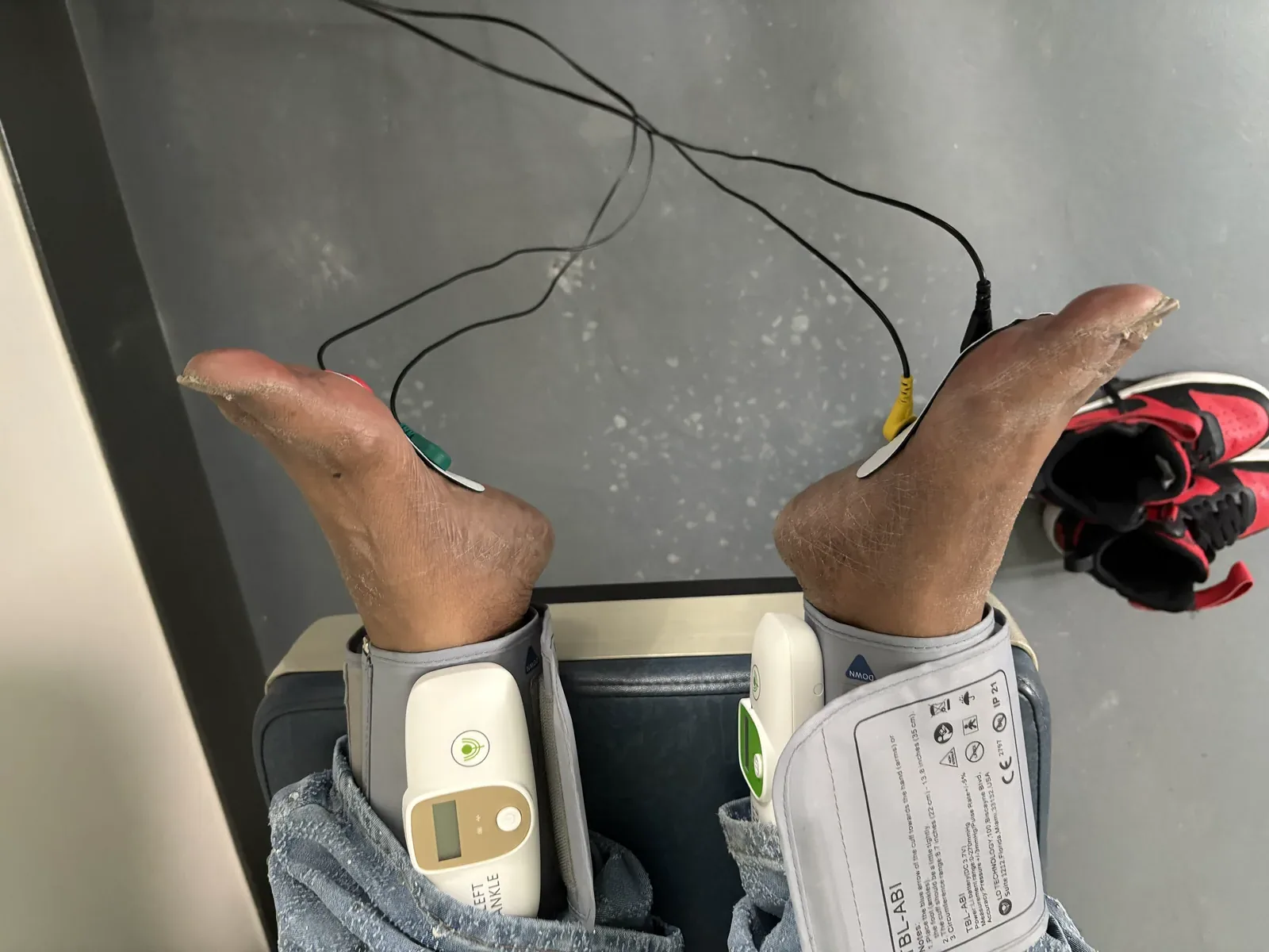
    Ankle blood pressure cuffs and sensors placed on a patient's feet during ankle-brachial index testing at Regenerve, 4477 Woodson Rd #104, St. Louis, MO 63134

    Testing that changes the plan

    Electrodiagnostic testing, performed on site, can characterize nerve involvement when symptoms are unclear or changing. Where perfusion is a question, vascular assessment looks at whether micro-circulation is part of why repair is slow. Both are worth doing when the answer would alter what you are doing at home.

    Movement, and why it belongs in a prevention plan

    Exercise works on the systems that decide how much stress your feet can take: insulin sensitivity and glucose handling, lipids, vascular function, and blood flow to the vasa nervorum that supply the nerve itself.

    The program structure used at Regenerve combines endurance work, sensorimotor and balance training, resistance work, and daily foot and ankle mobility. Details of the weekly structure and how to progress it without provoking flares are in exercises for peripheral neuropathy in the feet.

    What we do in clinic, and what it is not

    Medication that quiets the pain signal does not repair the nerve environment. The work in clinic is aimed at the terrain: restoring microvascular perfusion so oxygen reaches the nerve, reducing the metabolic pathways that produce glycation damage, and supplying the substrates nerves need, all under physician evaluation.

    Light-based therapies

    Class 4 photobiomodulation and class 3B cold laser are used as part of neuropathy care, alongside the metabolic work rather than instead of it. Whole-body infrared is also offered.

    Orthobiologic injections at Regenerve

    Where orthobiologic injections are considered, the driver-focused plan stays primary.

    When you find something

    This is where prevention is won or lost. Waiting for pain in a foot that cannot feel means waiting for the ulcer to declare itself some other way.

    • Stop walking on it. Keep weight and pressure off the area entirely.
    • Clean gently. Do not scrub broken skin or apply anything abrasive.
    • Call your clinician the same day for evaluation, not next week.

    Recurrence after a first ulcer is common enough that prevention has to continue after healing rather than stopping with it [2]. The routine is permanent.

    Frequently asked questions

    What does a daily foot check for neuropathy actually involve?

    Bright light, a mirror for the soles, and a look between every toe. You are watching for redness that does not fade, new callus, blisters, cracks, drainage or a new odor. It replaces the warning that protective sensation is no longer giving you. See peripheral neuropathy of the feet symptoms.

    Why do people with numb feet often end up in shoes that are too small?

    Because pressure is what still registers. Patients whose feet are numb may pick shoes half a size too small in order to feel that the shoe fits, which creates the pressure points that lead to skin breakdown [5]. Buy for measured length, not for how the shoe feels. See the hidden drivers of peripheral neuropathy.

    Do I need nerve testing to prevent a foot ulcer?

    No test substitutes for the daily check, but electrodiagnostic testing can clarify which fibers are involved when the pattern is unclear or changing, which in turn sharpens what you are preventing. See the neuropathy services offered at Regenerve.

    What if the problem is not diabetic neuropathy at all?

    It happens. Compression syndromes such as carpal tunnel and sciatica can produce numbness and burning that get filed under diabetic neuropathy, and the prevention plan differs. See carpal tunnel, sciatica and double crush.

    Find out what is driving it

    Prevention gets easier when you know which driver is doing the damage. The free five-question Nerve Damage Score at regenerve.com/assessment is the starting point, and it takes a few minutes.

    Sources

    1. Bragg S, Tucker Marrison S, Haley S, Diabetic Peripheral Neuropathy: Prevention and Treatment, American Family Physician, 2024;109(3):226–232, https://www.aafp.org/afp/2024/0300/diabetic-peripheral-neuropathy.
    2. Parveen K, Hussain MA, Anwar S, Elagib HM, Kausar MA, Comprehensive review on diabetic foot ulcers and neuropathy: treatment, prevention and management, World Journal of Diabetes, 2025;16(3):100329, https://pmc.ncbi.nlm.nih.gov/articles/PMC11885961/.
    3. Centers for Disease Control and Prevention, Diabetes and Your Feet, https://www.cdc.gov/diabetes/diabetes-complications/diabetes-and-your-feet.html.
    4. University of Michigan Medical School, The Burden of Diabetic Peripheral Neuropathy, https://medresearch.umich.edu/research-news/burden-diabetic-peripheral-neuropathy.
    5. Kowalick C, Advances in preventing and treating diabetic neuropathy, Magazines of the Schools at UT Health San Antonio, December 1, 2025, https://magazines.uthscsa.edu/schools/2025/12/01/advances-in-preventing-and-treating-diabetic-neuropathy/.
  • Duloxetine for Painful Diabetic Neuropathy: Evidence and Limits

    Duloxetine for Painful Diabetic Neuropathy: Evidence and Limits

    Duloxetine reduces pain more than placebo in painful diabetic peripheral neuropathy, and the size of that benefit is modest. In a 2023 systematic review and meta-analysis of 7 randomized controlled trials in adults with painful diabetic peripheral neuropathy, duloxetine was more efficacious than placebo for pain improvement, with a mean difference of -0.89 (95% confidence interval -1.09 to -0.69; P <.00001) [1].

    The limits sit in two places: whether you can tolerate it long enough for the benefit to matter, and whether the driver behind the nerve injury is still active while you take it.

    What the pooled trial evidence supports

    The same 2023 meta-analysis also found improvement on the Clinical Global Impression severity subscale, the Patient Global Impression of Improvement scale, and the European Quality of Life Instrument 5D version, all in patients with painful diabetic peripheral neuropathy [1].

    On dosing, the authors concluded that when a 60 mg dose is insufficient, 120 mg of duloxetine may improve symptoms [1]. That is an evidence statement, not a self-escalation instruction. Dose decisions belong with the prescribing clinician, particularly alongside other medications or with liver, blood pressure or bleeding considerations.

    What it does not do

    Duloxetine acts on pain signaling. It does not reduce glycation damage, improve delivery through the vasa nervorum, or correct a nutritional deficit. Where one of those is the active driver, the medication can help with the sensation while the underlying problem continues.

    Tolerability is where the evidence turns

    In the pooled trial data, severe adverse events were rare. Nausea, somnolence, dizziness, fatigue, constipation and decreased appetite were common, and approximately 12.6% of patients discontinued because of those common symptoms [1].

    A 2025 single-center study of 113 adults aged 25 to 65 with diabetic neuropathy, treated at a provincial hospital in Quetta, Pakistan, reported adverse effects in 32 patients (28.3%), most often nausea in 12 (10.6%), dizziness in 8 (7.1%) and somnolence in 7 (6.2%) [2]. Observational data cannot establish causation the way a randomized trial can, but it gives a sense of what tolerability looks like outside a trial protocol.

    Why the driver still matters

    Neuropathy is a category, not a diagnosis. Even when diabetes is clearly present, other contributors are frequently active at the same time, and they change what the rest of the plan should be.

    • Glycation and AGEs, which continue to act on nerve tissue regardless of what a pain medication is doing.
    • Functional B12 deficiency, which can be missed when a serum value in the normal range is treated as settled.
    • Gluten-related nerve injury, which can occur even with a negative celiac panel.
    • Medication effects, including agents that affect nutrient status and warrant monitoring.

    The clinical work at Regenerve is aimed at that layer: restoring microvascular perfusion so oxygen reaches the nerve, reducing the metabolic pathways producing glycation damage, and supplying the substrates nerves need, all under physician evaluation. That is the layer a pain medication does not reach.

    Testing that tells you whether the evidence applies to you

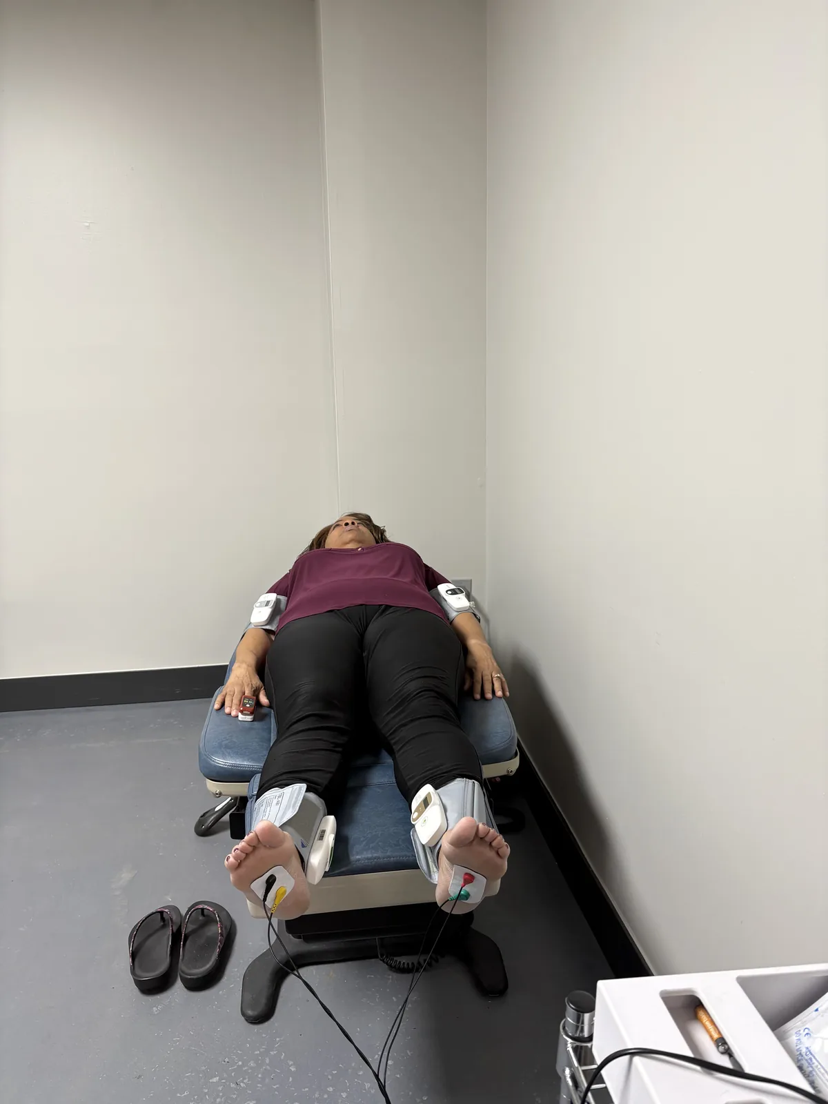
    Patient reclined for small-fiber and autonomic nerve testing, with sensor cuffs on both ankles and wrists, at Regenerve, 4477 Woodson Rd, St. Louis, MO 63134

    The trials above enrolled patients with painful diabetic peripheral neuropathy. Whether that describes your situation is a question the workup answers, not the diagnosis label on the chart.

    Electrodiagnostic testing

    EMG and nerve conduction studies, performed on site, characterize which fibers are involved and can separate a systemic neuropathy from a compression pattern. That matters most when symptoms suggest more than one mechanism at once.

    Balance testing is diagnostic only

    Videonystagmography, or VNG, measures inner-ear balance function. It clarifies whether a second system is contributing to unsteadiness. It is a test, not a treatment.

    Small fiber involvement

    Small fiber neuropathy can produce burning and sensory change that a single test will not always capture, which is one reason the evaluation focuses on the driver profile rather than on confirming the word neuropathy.

    What sits alongside medication

    None of this is framed as an alternative to duloxetine. Class 4 photobiomodulation and class 3B cold laser are used as part of neuropathy care, and whole-body infrared is also offered. Where gait and sensory feedback are affected, whole-body vibration may be part of the broader plan.

    For how these fit together for foot-predominant symptoms, see diabetic peripheral neuropathy treatment in St. Louis.

    Frequently asked questions

    How much pain relief does duloxetine actually provide?

    In a 2023 meta-analysis of 7 randomized controlled trials in adults with painful diabetic peripheral neuropathy, duloxetine beat placebo on pain improvement by a mean difference of -0.89 (95% CI -1.09 to -0.69; P <.00001) [1]. That is a real effect and a modest one, which is why it usually sits alongside driver-focused care rather than replacing it. See peripheral neuropathy treatments for the feet.

    What side effects most often make people stop?

    In the same pooled trial data, severe adverse events were rare, while nausea, somnolence, dizziness, fatigue, constipation and decreased appetite were common; about 12.6% of patients dropped out because of those common symptoms [1]. Tolerability, not efficacy, is usually the deciding factor. See small fiber neuropathy symptoms and testing.

    If 60 mg is not enough, does going to 120 mg help?

    The 2023 meta-analysis concluded that when a 60 mg dose is insufficient, 120 mg may improve symptoms in painful diabetic peripheral neuropathy [1]. That is a physician decision, and it depends on what else you take and on liver, blood pressure and bleeding risk. See why a negative celiac test does not close the question.

    What if my neuropathy is not only diabetic?

    Then the evidence base fits your situation less well, because these trials enrolled patients with painful diabetic peripheral neuropathy specifically. Nutritional, autoimmune, toxic and structural drivers can run alongside diabetes and change what the plan should prioritize. See why a normal B12 result can still miss a deficiency.

    Decide with a map, not a trial and error

    Whether duloxetine belongs in your plan is easier to answer once you know which driver is doing the most damage. The free five-question Nerve Damage Score at regenerve.com/assessment is where that conversation starts.

    Sources

    1. Wu CS, Huang YJ, Ko YC, Lee CH, Efficacy and safety of duloxetine in painful diabetic peripheral neuropathy: a systematic review and meta-analysis of randomized controlled trials, Systematic Reviews (BMC), 2023, doi:10.1186/s13643-023-02185-6, https://pmc.ncbi.nlm.nih.gov/articles/PMC10031998/.
    2. Osama M, Javaid M, Shahid B, Heema, Jamali AG, Anwar B, Mushtaq T, Efficacy of duloxetine in the management of diabetic neuropathy: a prospective observational cohort study, Cureus, 2025, doi:10.7759/cureus.82382, https://pmc.ncbi.nlm.nih.gov/articles/PMC12089739/.
  • Charcot Foot Warning Signs in Diabetic Neuropathy: What to Watch For

    Charcot Foot Warning Signs in Diabetic Neuropathy: What to Watch For

    The warning signs of Charcot foot are a feeling of heat or warmth in one foot, discoloration or redness, swelling, and foot pain — and Cleveland Clinic advises seeing a healthcare provider as soon as you notice any change in your feet.1 Charcot foot is rare, affecting less than 1% of all people with diabetes.1 The danger is not how common it is. The danger is the delay, because when neuropathy has reduced protective sensation, the foot can be under damage while reporting very little pain.

    Acute Charcot neuroarthropathy should be suspected in any patient 40 years or older with obesity and peripheral neuropathy who has an acutely swollen foot after minimal or no recalled trauma and who reports minimal to no pain, particularly when x-rays and laboratory markers of infection are normal.2 That description is worth reading twice, because it says the absence of pain is part of the picture rather than a reason to wait.

    Key takeaways

    • Compare both feet. Warmth on one side is the sign people most often notice first.
    • Do not wait for pain. Reduced sensation means the usual alarm may never sound.
    • Redness and swelling are not always infection. They still need same-week evaluation.
    • Delay has a cost. Delay in treatment results in the development of rigid foot and ankle deformities, which raises the risk of ulceration, infection and major lower-extremity amputation.2
    • The underlying neuropathy still needs a driver. Blood sugar is one contributor among several.

    What Charcot foot is, in plain language

    Charcot foot is a condition in which the bones and joints of the foot weaken and can change shape. It occurs in the setting of peripheral neuropathy, where reduced sensation allows repeated injury and inflammation to continue without the usual protective response of guarding a painful foot.

    Why neuropathy makes the warning signs easy to miss

    When sensation is reduced, pressure, friction or a minor twist can injure the foot without registering at the time. You may notice the swelling before you notice anything hurting, and that reversed order is exactly what makes this a looking problem rather than a feeling problem.

    This is why the symptoms people usually associate with neuropathy — burning at night, numbness, pins and needles — are not the ones to track here. Those describe the nerve. Warmth, swelling and color change describe the foot. Both matter, and they are checked differently. Our overview of peripheral neuropathy of the feet symptoms covers the nerve side.

    Bare feet of an older adult showing dry skin, thickened toenails and bunion deformity, the appearance often seen alongside peripheral neuropathy of the feet

    The early watch list

    These four early symptoms are the ones Cleveland Clinic lists, and all four can be checked at home without equipment.1

    • Heat or warmth, especially one foot noticeably warmer than the other
    • Discoloration or redness over the foot
    • Swelling, including a shoe that suddenly fits differently
    • Foot pain, which may be mild or absent when sensation is reduced

    How to check, in a way you will actually repeat

    1. Compare both feet at the same time of day, under the same conditions, using the back of your hand for temperature.
    2. Look at the soles with a mirror or a phone camera if you cannot lift the foot easily.
    3. Note whether a change followed extra walking, a new pair of shoes or a bump you barely registered.
    4. Write down what you see and the date. A photograph on the same day each week makes a slow change visible.
    Hand holding a round mirror to inspect the sole of a foot, the daily self-check recommended when protective sensation is reduced by peripheral neuropathy

    Why redness and swelling are not automatically infection

    A warm, red, swollen foot can look like cellulitis, and it is often treated as such first. In the setting of neuropathy, repetitive microtrauma can produce a very similar picture without infection being present, which is one reason the diagnosis is missed early.

    This is not a distinction to make at home. It is a reason to be evaluated rather than to wait and see which explanation fits better over the next two weeks.

    What happens when recognition is delayed

    A classic later sign is rocker-bottom foot, which occurs when the bones in the middle of the foot collapse and the arch drops.5 Once the shape of the foot has changed, pressure is distributed abnormally, and that abnormal pressure is what carries the risk forward into ulceration.

    The neuropathy underneath still needs a driver

    Charcot foot happens on top of peripheral neuropathy, and the neuropathy itself deserves the same scrutiny. In many patients diabetes is the dominant driver, but nutritional deficiency, autoimmune processes, toxic exposures, medications and structural compression can all be contributing alongside it.

    Identifying which of those is doing the most damage is the difference between a plan that reduces ongoing nerve injury and a plan that only manages symptoms. Our approach when diabetes is the primary driver is described on the diabetic peripheral neuropathy treatment page.

    Where Regenerve’s therapies do and do not fit

    Class 4 photobiomodulation, class 3B cold laser, whole-body infrared, and metabolic and nutritional care are used at Regenerve as part of a longer-term plan for the nerve environment. None of them is a response to an acutely warm, swollen foot. If Charcot warning signs appear, the correct next step is prompt clinical evaluation, not a therapy appointment.

    Orthobiologic injections (PRP and BMAC) are offered in selected situations.

    Where to get help in the St. Louis region

    Regenerve serves patients across the St. Louis region, Missouri and Illinois, including Edwardsville, Glen Carbon, Collinsville, Troy and Maryville. The clinic is at 4477 Woodson Rd #104, St. Louis, MO 63134, minutes from St. Louis Lambert International Airport.

    Call or text (314) 886-5902, or email info@regenerve.com. Hours are Monday through Thursday 9:00 to 16:00 and Friday 9:00 to 13:30. If a foot is acutely warm, red and swollen and you cannot be seen quickly, that is a reason to seek same-day care rather than to wait for a routine appointment.

    Your next step

    If you want to understand the neuropathy underneath the foot risk, the free five-question Nerve Damage Score identifies which driver category most likely applies to you, and your answers guide what we evaluate first.

    Frequently asked questions

    What are the first Charcot foot warning signs I should check for?

    A feeling of heat or warmth in one foot, especially when it is noticeably warmer than the other, along with swelling and discoloration or redness. Foot pain can also appear, but reduced sensation can mute it. Any of these needs prompt in-person evaluation; our guide to choosing a neuropathy specialist covers what to ask for.

    I have burning feet at night but no wound. Is that a Charcot warning sign?

    Burning at night is a nerve-pain symptom rather than a Charcot sign, and on its own it does not tell you whether the bones and joints are involved. Warmth on one side, swelling and color change are the findings that call for prompt evaluation. See burning feet and nerve pain.

    Does testing for neuropathy help with Charcot risk?

    Indirectly. Confirming that protective sensation is reduced tells you how much you have to rely on looking at the feet rather than feeling a problem, which is the whole basis of a daily check. Our on-site electrodiagnostic testing is described on the neuropathy services page.

    If I have diabetes, is blood sugar the only thing that raises my risk?

    No. Nutritional, autoimmune, toxic and structural contributors can coexist with diabetes and add to the nerve injury that makes a foot vulnerable. See the hidden drivers of peripheral neuropathy.

    Sources

    1. Cleveland Clinic, “Charcot Foot,” Cleveland Clinic health library, last updated June 30, 2023, https://my.clevelandclinic.org/health/diseases/15836-charcot-foot
    2. Marmolejo VS, Arnold JF, Ponticello M, Andersen CA, “Charcot Foot: Clinical Clues, Diagnostic Strategies, and Treatment Principles,” American Family Physician, May 1, 2018, https://www.aafp.org/afp/2018/0501/p594
    3. Wukich DK, Schaper NC, Gooday C, et al., on behalf of the International Working Group on the Diabetic Foot, “Guidelines on the diagnosis and treatment of active Charcot neuro-osteoarthropathy in persons with diabetes mellitus,” IWGDF, 2023, https://iwgdfguidelines.org/wp-content/uploads/2023/07/IWGDF-2023-08-Charcot-Guideline.pdf
    4. Rogers LC, Frykberg RG, Armstrong DG, et al., “The Charcot Foot in Diabetes,” Diabetes Care, 2011;34(9):2123–2129, https://diabetesjournals.org/care/article/34/9/2123/38608/The-Charcot-Foot-in-Diabetes
    5. MedlinePlus, “Charcot foot,” MedlinePlus Medical Encyclopedia, U.S. National Library of Medicine, review date November 7, 2024, https://medlineplus.gov/ency/article/007748.htm
    6. American Academy of Orthopaedic Surgeons, “Diabetic (Charcot) Foot,” OrthoInfo, https://www.orthoinfo.org/diseases–conditions/diabetic-charcot-foot/
    7. Collins AC, “Charcot Neuropathic Osteoarthropathy,” StatPearls, Treasure Island (FL): StatPearls Publishing, last updated January 31, 2026, https://www.ncbi.nlm.nih.gov/books/NBK470164/
  • Peripheral Neuropathy of the Feet: Symptoms, Patterns, and What They Mean

    Peripheral Neuropathy of the Feet: Symptoms, Patterns, and What They Mean

    Peripheral neuropathy in the feet does not announce itself. It arrives as something easy to explain away — socks that feel bunched when they are not, a floor that feels slightly wrong underfoot, toes that burn at night and are fine by morning. By the time most people describe it to a physician, the process has usually been running for years.

    The symptoms are worth reading carefully, because their pattern tells you a great deal about what is damaging the nerve and how far it has gone.

    Bare feet of an older adult showing dry skin, thickened toenails and bunion deformity, the appearance often seen alongside peripheral neuropathy of the feet

    The pattern matters more than any single symptom

    Metabolic neuropathy is length-dependent. It affects the longest nerve fibers first, which means it starts at the toes, spreads upward symmetrically on both sides, and only reaches the fingertips once it has climbed to roughly knee height. Clinicians call this a stocking-glove distribution, and it is the signature of a systemic process affecting every nerve at once rather than an injury to one nerve.1

    Two features of that pattern carry real diagnostic weight. It is symmetrical — both feet, roughly equally. And it ascends. Symptoms in one foot only, or symptoms that skip from a foot to a hand without covering the ground between, point somewhere else, often toward nerve compression or a double-crush pattern.

    Positive symptoms: what the nerve adds

    Damaged sensory nerves misfire, generating signals with no stimulus behind them. Patients describe these in remarkably consistent language:

    • Burning, often described as the feet being on fire, and often worst at night when there is nothing else competing for attention
    • Tingling or pins and needles that does not resolve when you move the foot
    • Electric or shooting pain arriving in bursts, sometimes without any trigger
    • Allodynia — pain from something that should not hurt, most classically the weight of a bedsheet on the toes
    • Deep aching in the arches or across the ball of the foot
    • Crawling, itching or wetness where nothing is touching the skin

    These are the symptoms that bring people in, and they are also the ones most likely to be treated in isolation. Relief matters. But a burning foot is a report about the nerve’s condition, and turning down the volume does not change what generated it.

    Negative symptoms: what the nerve stops delivering

    The quieter half of the picture is loss, and it is the more dangerous half:

    • Numbness, or a sense that the feet are wrapped in a layer of something
    • Reduced ability to feel temperature, which is why burns from hot water and heating pads are a genuine risk
    • Loss of position sense, so you no longer know where your feet are without looking at them
    • Unsteadiness, especially in the dark or on uneven ground — a very common first complaint
    • Weakness in the foot and ankle, sometimes showing up as tripping on curbs or scuffing a toe
    • Not noticing injury — the blister, the stone in the shoe, the crack in the heel

    That last item is the one that changes the stakes. When protective sensation is gone, the foot no longer reports damage, and a minor injury can progress without ever hurting. A 10-gram monofilament test identifies loss of protective sensation, and anyone who has it needs daily visual foot checks and well-fitted footwear as a standing routine.2

    Small fibers and large fibers give different symptoms

    Sensory nerves come in two calibers, and they fail differently. Small unmyelinated and thinly myelinated fibers carry pain, temperature and autonomic signals. Large myelinated fibers carry vibration, light touch and position sense.

    Small-fiber symptoms come first in most metabolic neuropathy: burning, pinprick pain, altered temperature sensation, and autonomic changes such as feet that no longer sweat, skin that has become dry and shiny, or color changes. Large-fiber symptoms arrive later: numbness, lost vibration sense, imbalance, and reduced or absent ankle reflexes.3

    This distinction has a practical consequence that catches a lot of patients. Standard nerve conduction studies measure large fibers. If your neuropathy is still predominantly small-fiber, those studies can come back normal while your feet are burning every night — and a normal result gets misread as “nothing is wrong.” It means the test looked at the wrong fibers. We cover this in detail in small fiber peripheral neuropathy symptoms and testing.

    Autonomic symptoms people rarely connect to their feet

    The same small fibers that carry pain also control sweating, blood vessel tone and internal organ function. When they are damaged, symptoms appear well beyond the feet: lightheadedness on standing, unexplained sweating changes, early fullness after meals, constipation or diarrhea, bladder changes, and erectile dysfunction. These are often reported to different specialists as separate problems. They frequently belong to one process.

    What the symptoms are usually telling you

    Peripheral neuropathy is not rare and it is not confined to people with diabetes. In a national sample of U.S. adults aged 40 and older, 13.5% had peripheral neuropathy — 27.0% of those with diabetes, and 11.6% of those without. In both groups it was independently associated with mortality over a median 13 years of follow-up.4 See diabetic peripheral neuropathy treatment in St. Louis.

    The metabolic connection runs deeper than a diabetes diagnosis. Among patients whose neuropathy had been labeled idiopathic, 34% turned out to have impaired glucose tolerance — about three times the rate in age-matched controls — and in many of them the fasting glucose and HbA1c were normal, with only a two-hour glucose tolerance test revealing the problem.5 A separate series found that even the normoglycemic patients with idiopathic neuropathy carried a heavy load of metabolic syndrome features, with lipid abnormalities especially prevalent.6

    So when the feet start burning, the useful question is not only “how bad is the nerve damage” but “what is the metabolic environment that produced it.” Those symptoms are frequently the first visible output of a systemic problem that has not yet been named. The mechanism is covered in how sugar damages nerves.

    When symptoms need attention sooner rather than later

    Most feet-first neuropathy develops over years. Some presentations should not wait:

    • Rapid progression over days or weeks
    • Marked weakness rather than mainly sensory symptoms
    • Symptoms that are clearly asymmetrical, or confined to one nerve’s territory
    • Symptoms beginning in the hands and feet at the same time
    • Any open wound, ulcer, unexplained swelling, redness or warmth in a numb foot
    • New bladder or bowel changes alongside the foot symptoms

    The last of these can indicate something above the peripheral nerve entirely and warrants prompt evaluation.

    How the symptoms get turned into a diagnosis

    “You have neuropathy” is a restatement of your symptoms, not an answer. A useful evaluation establishes three things: which fibers are affected, how far the damage has progressed, and what is driving it. That means a proper neurologic examination, electrodiagnostic testing where large-fiber involvement is in question, and a genuine search for the driver — metabolic, nutritional, autoimmune, toxic, pharmacologic or structural. At Regenerve, electrodiagnostic testing is performed on site. The full range of drivers worth ruling out is in the 11 hidden drivers of peripheral neuropathy, what treatment follows is in peripheral neuropathy treatments for the feet, and the non-drug options are graded in alternative medicine for peripheral neuropathy.

    Being evaluated in St. Louis

    Regenerve is at 4477 Woodson Rd #104, St. Louis, MO 63134, minutes from St. Louis Lambert International Airport, and is led by Dr. Gurpreet Singh Padda, MD, MBA, MHP. The practice serves the St. Louis region, Missouri and Illinois.

    Call or text (314) 886-5902, or email info@regenerve.com. Office hours are Monday through Thursday 9:00 to 16:00 and Friday 9:00 to 13:30. Details are on the contact page and the locations page.

    If you recognize your feet in this article, the Nerve Damage Score is a five-question assessment that produces a large-print report you can bring to any physician.

    Frequently asked questions

    What are the first symptoms of peripheral neuropathy in the feet?

    Usually tingling, burning or numbness starting in the toes of both feet, often worse at night, along with a vague sense of unsteadiness on uneven ground. See the hidden drivers of peripheral neuropathy.

    Why are my neuropathy symptoms worse at night?

    Fewer competing sensory inputs at rest make spontaneous nerve firing more noticeable, and skin temperature changes at night can amplify small-fiber symptoms. See small fiber neuropathy symptoms and testing.

    Does peripheral neuropathy always affect both feet?

    Metabolic neuropathy is characteristically symmetrical and affects both feet. Symptoms in one foot only suggest a different mechanism, such as nerve compression. See carpal tunnel, sciatica and double crush.

    My nerve test was normal but my feet still burn. Is that possible?

    Yes. Standard nerve conduction studies measure large fibers, and a small-fiber neuropathy can produce severe burning while those studies read normal. See small fiber neuropathy symptoms and testing.

    Is numbness in the feet more serious than pain?

    In terms of risk, yes. Numbness means the foot has stopped reporting injury, which is how minor wounds progress unnoticed. Daily visual foot checks become essential. See peripheral neuropathy treatments for the feet.

    Can I have neuropathy symptoms without diabetes?

    Yes. In a national U.S. sample, 11.6% of adults aged 40 and older without diabetes had peripheral neuropathy, and many idiopathic cases have impaired glucose tolerance or other metabolic syndrome features that routine testing missed. See how sugar damages nerves.

    Sources

    1. Tesfaye S, et al. Diabetic neuropathies: update on definitions, diagnostic criteria, estimation of severity, and treatments. Diabetes Care. 2010. PMID 20876709
    2. Boulton AJM, et al. Comprehensive foot examination and risk assessment: a report of the task force of the foot care interest group of the American Diabetes Association. Diabetes Care. 2008. PMID 18663232
    3. Terkelsen AJ, et al. The diagnostic challenge of small fibre neuropathy: clinical presentations, evaluations, and causes. The Lancet Neurology. 2017. PMID 29029847
    4. Hicks CW, Wang D, Matsushita K, Windham BG, Selvin E. Peripheral Neuropathy and All-Cause and Cardiovascular Mortality in U.S. Adults: A Prospective Cohort Study. Annals of Internal Medicine. 2021. PMID 33284680
    5. Singleton JR, Smith AG, Bromberg MB. Increased prevalence of impaired glucose tolerance in patients with painful sensory neuropathy. Diabetes Care. 2001. PMID 11473085
    6. Smith AG, Rose K, Singleton JR. Idiopathic neuropathy patients are at high risk for metabolic syndrome. Journal of the Neurological Sciences. 2008. PMID 18606421
  • Small Fiber Peripheral Neuropathy: Symptoms, Testing, and the Metabolic Cause Behind Most Cases

    Small Fiber Peripheral Neuropathy: Symptoms, Testing, and the Metabolic Cause Behind Most Cases

    Small fiber neuropathy is the diagnosis most often missed in people whose feet burn. The symptoms are severe, the standard nerve test is frequently normal, and the patient is sent home with the impression that nothing was found. Nothing was found because the test that was run does not look at the fibers that are damaged.

    This article covers what the symptoms actually are, how the condition is properly tested for, and why the cause — in a large share of cases — turns out to be metabolic.

    What “small fiber” means

    Peripheral sensory nerves come in two calibers. Large myelinated fibers carry vibration, light touch, position sense and the signals behind your ankle reflexes. Small fibers — thinly myelinated A-delta and unmyelinated C fibers — carry pain, temperature, and the autonomic traffic that controls sweating, blood vessel tone and internal organ function.1

    Small fibers are the most metabolically exposed part of the system. They are unmyelinated or barely myelinated, they terminate in the skin far from their cell bodies, and they depend on the smallest blood vessels in the body. When the metabolic environment deteriorates, they fail first — and they fail at the toes first, because that is where the longest ones end.

    The symptoms

    Sensory symptoms

    • Burning feet, the most characteristic complaint, typically worse at night and worse at rest
    • Pins and needles, prickling or tingling that does not resolve with movement
    • Sharp, stabbing or electric pain in bursts
    • Allodynia — the weight of bedding on the toes registering as pain
    • Altered temperature sensation, where cold feels burning or hot water cannot be judged reliably
    • A sense of walking on pebbles, sand, or bunched socks that are not there

    The distribution is symmetrical and length-dependent: toes first, then feet, then ascending the legs over months to years, with the hands involved only much later.

    Autonomic symptoms

    This is the half that rarely gets connected to the feet, because the symptoms appear elsewhere in the body and get reported to different specialists:

    • Reduced or absent sweating in the feet, leaving skin dry, cracked and shiny — or, less often, excessive sweating elsewhere
    • Lightheadedness on standing from impaired blood vessel constriction
    • Color and temperature changes in the feet — mottling, redness, or feet that feel cold to you and warm to the touch
    • Gastrointestinal changes: early fullness after eating, bloating, constipation or diarrhea
    • Bladder changes and erectile dysfunction
    • Dry eyes and dry mouth

    When burning feet and several of these appear together, small fiber neuropathy is a far better explanation than five unrelated diagnoses.1

    What is usually absent

    Pure small fiber neuropathy generally spares strength, vibration sense and reflexes. Normal ankle reflexes and normal strength do not argue against the diagnosis — they are what the diagnosis predicts. Once weakness, lost vibration sense and absent reflexes appear, large fibers have been recruited into the process as well.

    Why standard nerve testing comes back normal

    Nerve conduction studies and electromyography measure large myelinated fibers. Small fibers are, by definition, not what those studies record. A patient with pure small fiber neuropathy can have severe nightly burning and an entirely normal electrodiagnostic study.1

    This is worth being precise about, because a normal result is often reported to the patient as reassurance. It is not reassurance. It is a negative finding on a test aimed elsewhere. The correct interpretation is that large fibers are intact, which is useful information — it makes small-fiber-predominant disease more likely, not less.

    Electrodiagnostic testing still belongs in the workup. It rules out large-fiber involvement, identifies coexisting nerve compression, and establishes a baseline. At Regenerve it is performed on site, and it is always interpreted alongside the history, the examination and the search for a cause.

    Patient reclined for small-fiber and autonomic nerve testing, with sensor cuffs on both ankles and wrists, at Regenerve, 4477 Woodson Rd, St. Louis, MO 63134

    How small fiber neuropathy is confirmed

    The reference standard is a 3 mm punch skin biopsy from the distal leg, with intraepidermal nerve fiber density counted against age- and sex-matched normative values. The European Federation of Neurological Societies and Peripheral Nerve Society guideline rates this as a reliable and efficient method of confirming the diagnosis.2 It is a small procedure done under local anesthetic, and it produces a number that can be repeated later to measure change.

    Two other tests appear in the workup. Quantitative sensory testing measures thermal and pain thresholds, and quantitative sudomotor axon reflex testing measures sweat responses driven by the small autonomic fibers.1 Both add information; neither replaces the biopsy.

    The cause is metabolic more often than “idiopathic”

    A large proportion of small fiber neuropathy has historically been labeled idiopathic. Much of that label reflects incomplete testing.

    When 107 consecutive patients with idiopathic neuropathy were given a two-hour oral glucose tolerance test, 36 of them — 34% — had impaired glucose tolerance, nearly three times the rate in age-matched controls. Fasting glucose and HbA1c were frequently normal in those patients; the OGTT was the test that found it. The authors concluded that impaired glucose tolerance may cause or contribute to small-fiber neuropathy with a phenotype resembling the painful sensory neuropathy seen in diabetes.3

    The story does not end at glucose. In a series of 219 patients with idiopathic neuropathy, those with completely normal blood sugar carried the same elevated burden of metabolic syndrome features as those with impaired glucose tolerance, and had significantly higher total and LDL cholesterol and more abnormal HDL and triglycerides than a comparison group with diabetes but no neuropathy.4

    In other words: small fiber neuropathy tracks the metabolic terrain as a whole — glucose handling, lipids, blood pressure, central obesity and the microvascular supply — not any single laboratory value. Beyond the metabolic group, the differential includes autoimmune disease, B12 and other nutritional deficiencies, gluten-related nerve injury, thyroid disease, infections, toxic exposures, certain medications, and genetic sodium channel variants.1 The full list is in the 11 hidden drivers of peripheral neuropathy.

    Small fibers can regrow

    This is the part worth holding onto. Because small fiber density is measured in the skin, it can be measured again — and it moves.

    Thirty-two patients with impaired glucose tolerance and neuropathy received individualized diet and exercise counseling for one year. Intraepidermal nerve fiber density improved by 1.4 fibers per millimeter at the proximal biopsy site, and the improvement correlated with reduced neuropathic pain.5 Separately, a 10-week supervised aerobic and resistance exercise program in patients with diabetic peripheral neuropathy produced significant reductions in pain and neuropathic symptoms alongside increased intraepidermal nerve fiber branching on biopsy.6

    Both were small studies, and neither is a promise. But they demonstrate something that matters clinically: treating the metabolic environment produces measurable change in the nerve fibers themselves, not merely in how the patient rates their pain. That is the reasoning behind our approach, set out in the Regenerve Protocol. The training side is covered in exercises for peripheral neuropathy in the feet, and the supplement evidence — including alpha-lipoic acid, which has been studied in exactly this symptom set — is graded in alternative medicine for peripheral neuropathy.

    What a proper evaluation should include

    • A history and examination oriented to fiber type, not just to “does it hurt”
    • Electrodiagnostic testing to define large-fiber status and exclude compression
    • A two-hour oral glucose tolerance test, not a fasting glucose or HbA1c alone
    • A full lipid panel, read as part of the neuropathy workup rather than a separate cardiac issue
    • B12 with methylmalonic acid, thyroid studies, and screening for autoimmune and inflammatory causes
    • Skin biopsy for intraepidermal nerve fiber density where the diagnosis needs confirming or quantifying
    • A medication review, including statins and long-term metformin

    Being evaluated in St. Louis

    Regenerve is at 4477 Woodson Rd #104, St. Louis, MO 63134, minutes from St. Louis Lambert International Airport, and is led by Dr. Gurpreet Singh Padda, MD, MBA, MHP. The practice serves the St. Louis region, Missouri and Illinois.

    Call or text (314) 886-5902, or email info@regenerve.com. Office hours are Monday through Thursday 9:00 to 16:00 and Friday 9:00 to 13:30. Details are on the contact page and the locations page.

    If a nerve test came back normal and your feet still burn, the Nerve Damage Score is a five-question assessment that produces a large-print report you can bring to any physician.

    Frequently asked questions

    What are the symptoms of small fiber peripheral neuropathy?

    Burning feet that are worse at night, prickling and stabbing pain, pain from light touch such as bedding, altered temperature sensation, and autonomic symptoms including reduced sweating in the feet, lightheadedness on standing and gastrointestinal changes. See peripheral neuropathy of the feet symptoms.

    Is “small fibre neuropathy” the same as small fiber neuropathy?

    Yes. “Small fibre” is the British spelling of the same condition; the diagnosis, testing and treatment are identical. See the hidden drivers of peripheral neuropathy.

    Can small fiber neuropathy show up with a normal EMG and nerve conduction study?

    Yes, and it commonly does. Those studies measure large myelinated fibers, so a pure small fiber neuropathy can produce severe symptoms with a normal result. See which specialist you should see.

    How is small fiber neuropathy diagnosed?

    By a 3 mm punch skin biopsy from the distal leg with intraepidermal nerve fiber density counted against normative values, supported by quantitative sensory and sudomotor testing. See the Regenerve Protocol.

    What causes small fiber neuropathy?

    Metabolic causes lead. About a third of patients labeled idiopathic have impaired glucose tolerance that fasting glucose and HbA1c missed, and even normoglycemic patients carry a high burden of metabolic syndrome features, particularly abnormal lipids. See how sugar damages nerves.

    Can small nerve fibers grow back?

    They can. Diet and exercise counseling over one year increased intraepidermal nerve fiber density on skin biopsy, and the increase correlated with less neuropathic pain. Recovery is partial and slow, and no one can promise a cure. See exercises for peripheral neuropathy in the feet.

    Sources

    1. Terkelsen AJ, et al. The diagnostic challenge of small fibre neuropathy: clinical presentations, evaluations, and causes. The Lancet Neurology. 2017. PMID 29029847
    2. Lauria G, et al. European Federation of Neurological Societies/Peripheral Nerve Society Guideline on the use of skin biopsy in the diagnosis of small fibre neuropathy. European Journal of Neurology. 2010. PMID 20642627
    3. Singleton JR, Smith AG, Bromberg MB. Increased prevalence of impaired glucose tolerance in patients with painful sensory neuropathy. Diabetes Care. 2001. PMID 11473085
    4. Smith AG, Rose K, Singleton JR. Idiopathic neuropathy patients are at high risk for metabolic syndrome. Journal of the Neurological Sciences. 2008. PMID 18606421
    5. Smith AG, et al. Lifestyle intervention for pre-diabetic neuropathy. Diabetes Care. 2006. PMID 16732011
    6. Kluding PM, et al. The effect of exercise on neuropathic symptoms, nerve function, and cutaneous innervation in people with diabetic peripheral neuropathy. Journal of Diabetes and its Complications. 2012. PMID 22717465
  • Peripheral Neuropathy: The 11 Hidden Drivers Your Doctor Missed

    Peripheral Neuropathy: The 11 Hidden Drivers Your Doctor Missed

    When a workup comes back inconclusive, many patients are told their neuropathy is “idiopathic” — medical language for cause unknown. But in a large share of these cases, the cause isn’t truly unknown; it simply hasn’t been looked for thoroughly enough. Idiopathic is too often a clinical surrender rather than a diagnosis. This article lays out the systematic investigation Dr. Padda uses to hunt down the real driver of nerve damage, organized into three domains — metabolic, toxic, and mechanical — because a nerve can die from many directions, and finding which one is the difference between managing symptoms forever and actually changing course.

    “Idiopathic” is often incomplete, not unsolvable

    Studies of unexplained peripheral neuropathy consistently show that when patients undergo a structured, expanded evaluation, an identifiable cause emerges in a substantial proportion of cases previously labeled idiopathic. The American Academy of Neurology’s evaluation guidance for distal symmetric polyneuropathy emphasizes a tiered laboratory workup precisely because targeted testing changes management. The point is not that every case has an easy answer — some remain genuinely unexplained — but that the label should be earned only after a real search.

    The three-domain framework below is a way to organize that search so nothing obvious gets skipped.

    Domain one: metabolic drivers

    Metabolic problems are the most common and most treatable causes of nerve damage. They share a final common pathway — energy failure and chemical injury inside the nerve.

    1. Blood sugar and glycation. Diabetes is the leading cause of neuropathy worldwide, but the damage begins earlier than most people realize. Research from the University of Utah (Drs. Smith and Singleton) helped establish that even prediabetes — impaired glucose tolerance that never reaches the diabetes threshold — is associated with small-fiber neuropathy. Chronically elevated glucose bonds to nerve proteins to form advanced glycation end-products through the Maillard reaction, first described by Louis-Camille Maillard in 1912. A standard fasting glucose can miss this entirely; markers like HbA1c and a glucose tolerance test reveal far more. (Explored in depth in the glycation and blood-sugar articles.)

    2. Intracellular nutritional deficiency. Nerves are cofactor-hungry. Deficiencies of B1 (thiamine), B6, B12, folate, magnesium, and omega-3 fatty acids each impair nerve function and mitochondrial energy production. Crucially, blood levels can look “normal” while the tissue is starved — which is why functional testing matters (see driver 3).

    3. Functional B12 deficiency. A serum B12 in the normal range does not rule out a cellular deficiency. Functional markers — methylmalonic acid (MMA) and homocysteine — rise when B12 is functionally inadequate at the tissue level, catching deficiencies a standard B12 test misses. Untreated, B12 deficiency causes a characteristic neuropathy (and can damage the spinal cord), yet it is eminently correctable. (Covered fully in the B12 article.)

    Domain two: toxic drivers

    If metabolic drivers are about deprivation, toxic drivers are about poisoning — substances that damage nerves directly or by depleting the body’s defenses.

    4. Bioaccumulated heavy metals. Lead, arsenic, mercury, and thallium disrupt essential enzymes and deplete glutathione, the body’s master antioxidant. Exposure is often occupational or environmental and accumulates silently over years. Testing should be guided by a genuine exposure history — and, importantly, patients should be steered away from unproven or aggressive “detox” schemes, which can do more harm than good. (See the heavy-metals article.)

    5. Mold and mycotoxins. Toxins produced by mold in water-damaged buildings can impair mitochondrial function and drive neuroinflammation, sometimes presenting as neuropathy paired with profound fatigue. This is a genuinely debated clinical area, and it deserves careful, evidence-aware evaluation rather than either dismissal or overdiagnosis. (See the mycotoxin article.)

    6. Neurotoxic medications. Some of the most commonly prescribed drugs can quietly undermine nerve health. Statins can deplete CoQ10; metformin — one of the most-prescribed diabetes drugs — can cause B12 deficiency over time; certain chemotherapy agents and antibiotics are directly neurotoxic. The answer is rarely to stop a needed medication, but to monitor and replete the nutrients they affect. (See the statins-and-metformin article.)

    7. Alcohol. Alcohol is a double hit: acetaldehyde and alcohol itself are directly toxic to nerves, and heavy use depletes thiamine and other B vitamins. Recovery depends on reducing exposure and repleting nutrients — but abrupt cessation in someone alcohol-dependent carries its own medical risks and should be handled with clinical guidance. (See the alcohol article.)

    Domain three: mechanical and immune drivers

    The final domain covers physical and immune-mediated injury — nerves crushed, choked, or caught in the crossfire of the body’s own defenses.

    8. Autoimmune disease. Conditions like lupus, rheumatoid arthritis, and Sjögren’s damage nerves through vasculitis (inflammation of the small vessels feeding the nerve) and direct antibody attack. Here the neuropathy is collateral damage from a systemic process, so treatment requires controlling the underlying disease and addressing the nerve. (See the autoimmune article.)

    9. Gluten sensitivity. Even without celiac disease, gluten can trigger a neurological immune response — antibodies against transglutaminase-6 have been linked to neuropathy and cerebellar ataxia in work led by Dr. Marios Hadjivassiliou. A negative celiac test does not rule this out. (See the two gluten articles.)

    10. Chronic stealth infections. Certain infections hide in nervous tissue. The varicella-zoster (shingles) virus resides in the dorsal root ganglia and can reactivate to cause postherpetic neuralgia; Lyme and other pathogens can also produce neuropathy. (See the shingles article.)

    11. Mechanical compression. Sometimes it isn’t a metabolic disease at all — it’s a pinched nerve. Carpal tunnel syndrome, radiculopathy (sciatica), and other entrapments injure nerves through ischemia and focal demyelination. The concept of double crush syndrome, described by Upton and McComas in 1973, explains why a metabolically stressed nerve is more vulnerable to a second, mechanical injury — meaning compression and metabolic disease often compound each other. (See the compression article.)

    Why one patient often has several drivers at once

    These categories are not mutually exclusive. A person with prediabetes, a statin prescription, and a compressed nerve at the wrist may have three simultaneous drivers, each amplifying the others. This is the practical reason a single-cause mindset fails: the workup has to be broad enough to catch combinations, and the treatment plan has to address all the active contributors, not just the most obvious one.

    What a thorough workup looks like

    A genuine root-cause investigation typically includes a detailed history (occupation, exposures, diet, alcohol, medications, family history), an expanded metabolic panel (glucose tolerance and HbA1c, not just fasting glucose), functional nutrient testing (MMA and homocysteine, thiamine, magnesium), targeted autoimmune and infectious testing when the history points that way, and — where relevant — nerve conduction studies or skin biopsy to characterize the fiber types involved. The aim is to convert “idiopathic” into a named, addressable cause whenever the evidence allows.

    Frequently asked questions

    My tests were normal — does that mean there’s no cause?

    Not necessarily. Standard panels can miss functional deficiencies (like tissue-level B12), early glucose dysregulation, and toxic exposures. A broader, targeted workup often uncovers a driver that routine testing skips.

    That is the subject of Mystery Neuropathy and Exhaustion? The Mold and Mycotoxin Question.

    Can more than one thing be causing my neuropathy?

    Yes, and it’s common. Multiple drivers frequently coexist and compound one another, which is why a complete evaluation matters.

    This is set out in The Sugar That Caramelizes Your Nerves: How Glucose Damages Your Nervous System.

    Is it too late if I’ve had neuropathy for years?

    Identifying and removing an active driver can slow or halt progression at any stage, and some function may recover. Earlier is better, but a workup is worthwhile regardless of how long symptoms have been present.

    There is more on this in Your “Normal” B12 May Be Lying: The Hidden Deficiency Driving Your Neuropathy.

    Should I try a detox for heavy metals?

    Only under medical guidance and only if testing and history justify it. Unproven chelation and “detox” protocols carry real risks and should not be self-administered.

    Mystery Neuropathy and Exhaustion? The Mold and Mycotoxin Question explains what that looks like.

    Key takeaways

    • “Idiopathic” often means the search was incomplete, not that no cause exists.
    • Nerve damage arises from metabolic, toxic, and mechanical drivers — often several at once.
    • Standard labs miss a lot: functional B12 markers, glucose tolerance, and exposure-guided testing reveal more.
    • Double crush syndrome shows how metabolic and mechanical injuries compound each other.
    • The goal of the workup is to convert an unexplained neuropathy into a named, treatable one.

    This article is educational and is not a substitute for evaluation, diagnosis, or treatment by a physician. Individual results vary. Do not start, stop, or change any medication or treatment without consulting your physician. Take the free Nerve Damage Score or call/text (314) 886-5902 to begin a root-cause evaluation.

    References

    1. England JD, Gronseth GS, Franklin G, et al. Distal symmetric polyneuropathy: a definition for clinical research (AAN/AANEM/AAPM&R). Neurology. 2005; and the AAN evaluation guidance, Neurology. 2009.
    2. Smith AG, Singleton JR. Impaired glucose tolerance and neuropathy. Neurologist / Diabetes Care (University of Utah body of work).
    3. Maillard LC. Action des acides aminés sur les sucres. C R Acad Sci. 1912.
    4. Brownlee M. Biochemistry and molecular cell biology of diabetic complications. Nature. 2001;414:813–820.
    5. Hadjivassiliou M, et al. Transglutaminase-6 antibodies and gluten-related neurological dysfunction. Neurology / Ann Neurol.
    6. Upton ARM, McComas AJ. The double crush in nerve-entrapment syndromes. Lancet. 1973;2(7825):359–362.

    Note: match each reference to a specific, current source at publication; several point to bodies of work rather than a single paper.

    Find out what is driving your nerve pain

    The free, five-question Nerve Damage Score takes about two minutes and tells you which terrain failure is most likely behind your symptoms.

    Get My Free Nerve Damage Score

    Or call or text (314) 886-5902.

  • The Sugar That Caramelizes Your Nerves: How Glucose Damages Your Nervous System

    The Sugar That Caramelizes Your Nerves: How Glucose Damages Your Nervous System

    The same chemistry that browns a steak in a hot pan and gives bread its crust is happening, slowly and silently, inside the body of anyone with chronically elevated blood sugar. It is called the Maillard reaction, and when it plays out on the proteins of your nervous system, the result is stiffened, short-circuited, and eventually dying nerves. This article explains — in plain terms but with real biochemistry — how glucose damages nerves through glycation, why it produces the burning feet of diabetic neuropathy, and why a single fasting glucose test can miss the process entirely.

    The Maillard reaction: caramelizing from the inside

    In 1912, the French chemist Louis-Camille Maillard described what happens when sugars react with proteins: they bond together and form new, brown, rigid compounds. In cooking, that reaction creates flavor and color. In the body, the same reaction runs on your own tissues whenever glucose is abundant. Glucose molecules latch onto proteins — including the structural and functional proteins of nerves — and, through a series of steps, form stable end-products.

    These are called Advanced Glycation End-products, or AGEs — an apt acronym, because the process is a form of accelerated aging of the tissue. Once formed, AGEs are hard to remove, and they accumulate over years of elevated blood sugar.

    Three ways AGEs injure nerves

    Glycation harms nerves through several converging mechanisms.

    1. Structural cross-linking. AGEs bind proteins to one another, cross-linking them into stiff, dysfunctional complexes. In a nerve, this degrades the delicate architecture required to conduct signals and to maintain the insulating myelin sheath. Cross-linked proteins in the walls of the tiny blood vessels feeding the nerve also stiffen those vessels, choking the nerve’s blood supply.

    2. Inflammatory ignition through RAGE. AGEs are not just inert debris. They bind to a specific receptor called RAGE (the receptor for advanced glycation end-products) on the surface of cells. Activating RAGE switches on inflammatory signaling cascades, flooding the tissue with inflammatory mediators and oxidative stress. The nerve is effectively set on a low, chronic inflammatory fire.

    3. Oxidative stress and the polyol pathway. When glucose is abundant, some of it is shunted through the polyol pathway, where the enzyme aldose reductase converts glucose to sorbitol. This process consumes NADPH — the same molecule the cell needs to regenerate glutathione, its master antioxidant. The nerve is thus hit twice: sorbitol accumulates and draws water into the cell, while the antioxidant defense is depleted just as oxidative stress is rising.

    The unifying mechanism: mitochondrial overload

    How do these pathways connect? The landmark work of Dr. Michael Brownlee, published in Nature in 2001, proposed a unifying explanation: excess glucose overloads the mitochondria — the cell’s power plants — causing them to overproduce a damaging molecule called superoxide. That single upstream event, Brownlee argued, switches on the glycation, polyol, and inflammatory pathways together. In other words, mitochondrial overload is the common root, and AGEs, sorbitol accumulation, and RAGE-driven inflammation are its branches. This is why effective treatment has to consider mitochondrial health, not just blood sugar numbers in isolation.

    Why the feet burn first

    Nerves signal by maintaining a precise electrical and chemical environment along their length. As glycation stiffens their structure, inflammation irritates them, and oxidative stress and poor blood flow starve them of energy, the fibers begin to misfire — generating the burning, tingling, and electric sensations of neuropathy — and then to die back. Because the process is length-dependent, the longest nerves, which reach the feet, are affected first. That is the biochemical reason diabetic neuropathy characteristically begins in the toes and moves upward.

    Beyond the feet: the brain connection

    The reach of glucose-driven damage does not stop at the peripheral nerves. Research by Dr. Suzanne de la Monte at Brown University introduced the concept of Alzheimer’s disease as, in part, a metabolic disorder of brain insulin signaling — sometimes called “type 3 diabetes.” The same terrain of glycation, inflammation, and insulin resistance that injures peripheral nerves also appears to affect the brain, which is one reason chronic high blood sugar is associated with cognitive symptoms as well as neuropathy.

    Why a single glucose test isn’t enough

    A one-time fasting glucose is a snapshot; glycation is a movie. Because AGE formation depends on cumulative sugar exposure over time, better windows into the process include HbA1c (which reflects average glucose over roughly three months and is itself a glycated protein), a glucose tolerance test (which can reveal impaired glucose handling that fasting numbers miss), and markers of the downstream damage. Catching dysregulation at the prediabetes stage — before a formal diabetes diagnosis — matters, because nerve damage can begin there.

    What this means for treatment

    The biochemistry points directly at the strategy: reduce the ongoing glucose exposure driving glycation, support the antioxidant defenses (glutathione and its cofactors) that the polyol pathway depletes, and restore mitochondrial function so the upstream overload eases. This is the rationale behind a terrain-focused approach that pairs glycemic control with targeted metabolic and mitochondrial support, rather than relying only on drugs that mask the resulting pain.

    Frequently asked questions

    Can nerve damage from high blood sugar be undone?

    Some can, especially when caught early and when the underlying glucose exposure and oxidative stress are corrected. Established damage may only partly recover, so the priority becomes halting progression. Individual results vary.

    My fasting glucose is normal — am I in the clear?

    Not necessarily. HbA1c and a glucose tolerance test reveal patterns a single fasting number misses, and nerve damage can begin at the prediabetes stage.

    What are AGEs, in one sentence?

    Advanced glycation end-products are stiff, damaging compounds formed when sugar bonds to your proteins — the body’s internal version of caramelization.

    Does this affect anything besides my feet?

    Yes. The same metabolic terrain is linked to blood-vessel disease and to cognitive effects; research describes an insulin-resistance component of brain disease sometimes called “type 3 diabetes.”

    Key takeaways

    • The Maillard reaction bonds glucose to your proteins, forming stiff, damaging AGEs.
    • AGEs harm nerves by cross-linking structure, activating RAGE-driven inflammation, and driving oxidative stress via the polyol pathway.
    • Brownlee’s work identifies mitochondrial overload as the unifying upstream event.
    • Damage is length-dependent, so the feet are affected first.
    • HbA1c and glucose tolerance testing reveal the process better than a single fasting glucose.

    This article is educational and is not a substitute for evaluation, diagnosis, or treatment by a physician. Individual results vary. Do not start, stop, or change any medication without consulting your physician. Take the free Nerve Damage Score or call/text (314) 886-5902.

    References

    1. Maillard LC. Action des acides aminés sur les sucres; formation des mélanoïdines par voie méthodique. C R Acad Sci. 1912;154:66–68.
    2. Brownlee M. Biochemistry and molecular cell biology of diabetic complications. Nature. 2001;414:813–820.
    3. Vlassara H, Uribarri J. Advanced glycation end products (AGEs) and diabetes. Curr Diab Rep. 2014.
    4. de la Monte SM, Wands JR. Alzheimer’s disease is type 3 diabetes—evidence reviewed. J Diabetes Sci Technol. 2008;2(6):1101–1113.
    5. Oates PJ. Polyol pathway and diabetic peripheral neuropathy. Int Rev Neurobiol. 2002.

    Find out what is driving your nerve pain

    The free, five-question Nerve Damage Score takes about two minutes and tells you which terrain failure is most likely behind your symptoms.

    Get My Free Nerve Damage Score

    Or call or text (314) 886-5902.

  • The Cruel Irony: How Statins and Metformin Can Affect Nerve Health

    The Cruel Irony: How Statins and Metformin Can Affect Nerve Health

    There is an uncomfortable irony at the heart of this topic: two of the most widely prescribed medications in the world — statins for cholesterol and metformin for diabetes — can, in some people, quietly deplete the very nutrients that nerves depend on. This does not mean these drugs are villains; they prevent heart attacks, strokes, and the progression of diabetes, and for most people the benefits are substantial. The point of this article is not to frighten anyone off necessary treatment, but to explain a real, manageable interaction so that the nutrients can be monitored and replaced. The answer here is repletion and monitoring, not abandoning needed medication.

    Metformin and vitamin B12

    Metformin is a cornerstone of diabetes care, and a good one. But a well-documented side effect of long-term use is impaired absorption of vitamin B12. Over years, this can produce a functional or overt B12 deficiency — and because B12 deficiency itself causes peripheral neuropathy, the result can be a patient whose diabetes drug is contributing to the very nerve symptoms being blamed entirely on their diabetes.

    This is one of the clearer, better-studied drug–nutrient interactions, and it has a clean solution: periodic monitoring of B12 status in people on long-term metformin, ideally using functional markers like methylmalonic acid and homocysteine rather than serum B12 alone (as discussed in the B12 article), and repletion when needed. Crucially, this is done without stopping metformin — the drug continues to do its job while the B12 is replaced.

    Statins and CoQ10

    Statins lower cholesterol by inhibiting an enzyme (HMG-CoA reductase) early in the cholesterol synthesis pathway. That same pathway also produces coenzyme Q10 (CoQ10), a molecule essential for mitochondrial energy production. By design, statins can therefore lower CoQ10 levels — and since nerves are metabolically demanding, the theoretical concern is that reduced CoQ10 could affect nerve energy metabolism.

    Honesty is important here, because this is a more debated area than the metformin–B12 link. The CoQ10-depletion mechanism is real. Whether statins meaningfully cause peripheral neuropathy has been studied and remains genuinely contested — some observational studies have suggested an association, while others have not confirmed a clear causal link, and any absolute risk appears small. The most defensible position is measured: statin-associated muscle symptoms are well recognized, a neuropathy association is possible but not firmly established, and the benefits of statins for cardiovascular protection are strong and well proven.

    The sensible framework: don’t stop — monitor and support

    Put together, these interactions call for a specific, non-alarmist approach.

    First, do not stop a statin or metformin on your own. Discontinuing a needed cardiovascular or diabetes medication carries real, sometimes serious, risks that typically outweigh the nutrient concerns.

    Second, monitor. In people on long-term metformin, check B12 status functionally and periodically. In people on statins with new muscle or nerve symptoms, discuss them with the prescribing clinician.

    Third, replete intelligently. Where B12 is low, replace it. Where CoQ10 supplementation is being considered for statin-related symptoms, that is a reasonable, low-risk conversation to have with a physician, recognizing that the evidence for benefit is mixed.

    Fourth, look at the whole picture. A person with diabetes on metformin and a statin may have several simultaneous contributors to their neuropathy — the diabetes itself, a metformin-related B12 deficiency, and possibly others. Sorting out how much each contributes is exactly the work of a root-cause evaluation.

    Other medications worth knowing about

    Metformin and statins are the headliners, but they are not alone. Certain chemotherapy agents are directly neurotoxic and a well-known cause of neuropathy; some antibiotics (including certain fluoroquinolones and long-term use of others) and a handful of other drugs can affect nerves as well. The recurring principle applies: the goal is informed monitoring and, where possible, mitigation — in partnership with the prescriber — rather than reflexive discontinuation.

    Frequently asked questions

    Should I stop my metformin or statin if I have neuropathy?

    No. Never stop these on your own — the risks of doing so are significant. Instead, ask your physician to check the relevant nutrient status and evaluate your symptoms.

    Does metformin definitely cause B12 deficiency?

    Long-term metformin use is a well-documented cause of impaired B12 absorption in a meaningful subset of users. It is manageable by monitoring and repletion while continuing the drug.

    Do statins cause neuropathy?

    The CoQ10-depletion mechanism is real, but whether statins meaningfully cause peripheral neuropathy is debated and any risk appears small. Muscle symptoms are the better-recognized statin side effect. Discuss new symptoms with your prescriber.

    Is it safe to take CoQ10 with a statin?

    CoQ10 is generally low-risk, and some people take it for statin-related symptoms, though the evidence for benefit is mixed. Discuss it with your physician.

    Key takeaways

    • Metformin can impair B12 absorption over time, contributing to neuropathy — a well-documented, manageable interaction.
    • Statins can lower CoQ10; a neuropathy link is possible but debated, and any risk appears small.
    • The answer is monitoring and repletion, never stopping needed medication on your own.
    • Functional B12 testing catches deficiencies a standard test misses in metformin users.
    • Multiple contributors often coexist, which is why the full picture matters.

    This article is educational and is not a substitute for evaluation, diagnosis, or treatment by a physician. Individual results vary. Do not start, stop, or change any medication without consulting your physician. Take the free Nerve Damage Score or call/text (314) 886-5902.

    References

    1. Aroda VR, Edelstein SL, Goldberg RB, et al. Long-term metformin use and vitamin B12 deficiency in the Diabetes Prevention Program Outcomes Study. J Clin Endocrinol Metab. 2016;101(4):1754–1761.
    2. Infante M, et al. Metformin, vitamin B12 deficiency and peripheral neuropathy (review). Endocrine / review.
    3. Marcoff L, Thompson PD. The role of coenzyme Q10 in statin-associated myopathy. J Am Coll Cardiol. 2007;49:2231–2237.
    4. Emad M, et al. Statins and peripheral neuropathy — evidence and controversy (review).

    Find out what is driving your nerve pain

    The free, five-question Nerve Damage Score takes about two minutes and tells you which terrain failure is most likely behind your symptoms.

    Get My Free Nerve Damage Score

    Or call or text (314) 886-5902.