
Category: Autoimmune, Infectious & Structural

  • CIDP vs. Length-Dependent Peripheral Neuropathy: How They Differ

    CIDP vs. Length-Dependent Peripheral Neuropathy: How They Differ

    CIDP and length-dependent peripheral neuropathy differ in mechanism, distribution and timeline. CIDP — chronic inflammatory demyelinating polyradiculoneuropathy — is immune-mediated inflammation of the nerve roots and peripheral nerve, and its symptoms must persist for at least 8 weeks to establish the diagnosis [1]. Length-dependent neuropathy instead follows the length of the nerve fiber, so assessment starts distally at the dorsal aspect of the great toe on both sides and moves upward [3].

    That difference is not academic. It changes which pattern your clinician is looking for, which tests are worth running, and in what order.

    What CIDP is

    CIDP is immune-mediated, involving inflammation of both nerve roots and peripheral nerve [1]. Its pathology is segmental demyelination and remyelination, and repeated cycles of that process produce the concentrically arranged Schwann cells known as onion bulbs [1].

    Because nerve roots are involved, the weakness does not stay in the feet. The typical presentation is symmetric progressive proximal and distal muscle weakness with distal numbness evolving over at least eight weeks [2].

    Course

    CIDP can be monophasic, relapsing or progressive, with roughly one-third of patients experiencing a relapsing-remitting pattern and others showing steady progression [1]. A course that flares and settles is one of the features that raises the question.

    Which sensations are affected

    CIDP particularly affects position and vibration sense more than pain and temperature, which reflects predominant large-fiber involvement [1]. Patients often describe unsteadiness and a sense that they cannot tell where their feet are, rather than burning.

    What length-dependent neuropathy is

    Length-dependent neuropathy is the pattern in which the longest nerve fibers are affected first, so symptoms begin in the toes and progress upward. In diabetes, the American Diabetes Association describes assessment as following the typical distal symmetric polyneuropathy pattern, starting distally at the dorsal aspect of the great toe on both sides and moving proximally [3].

    Which sensations are affected

    The most common early symptoms come from small-fiber involvement and include pain and dysesthesias — unpleasant burning sensations — described as burning, lancinating, tingling or shooting [3]. Large-fiber involvement causes numbness, tingling without pain, and loss of protective sensation, which marks more advanced neuropathy and raises the risk of foot ulceration [3].

    What that pattern means in practice, and how it is examined, is covered in peripheral neuropathy of the feet, symptoms and patterns.
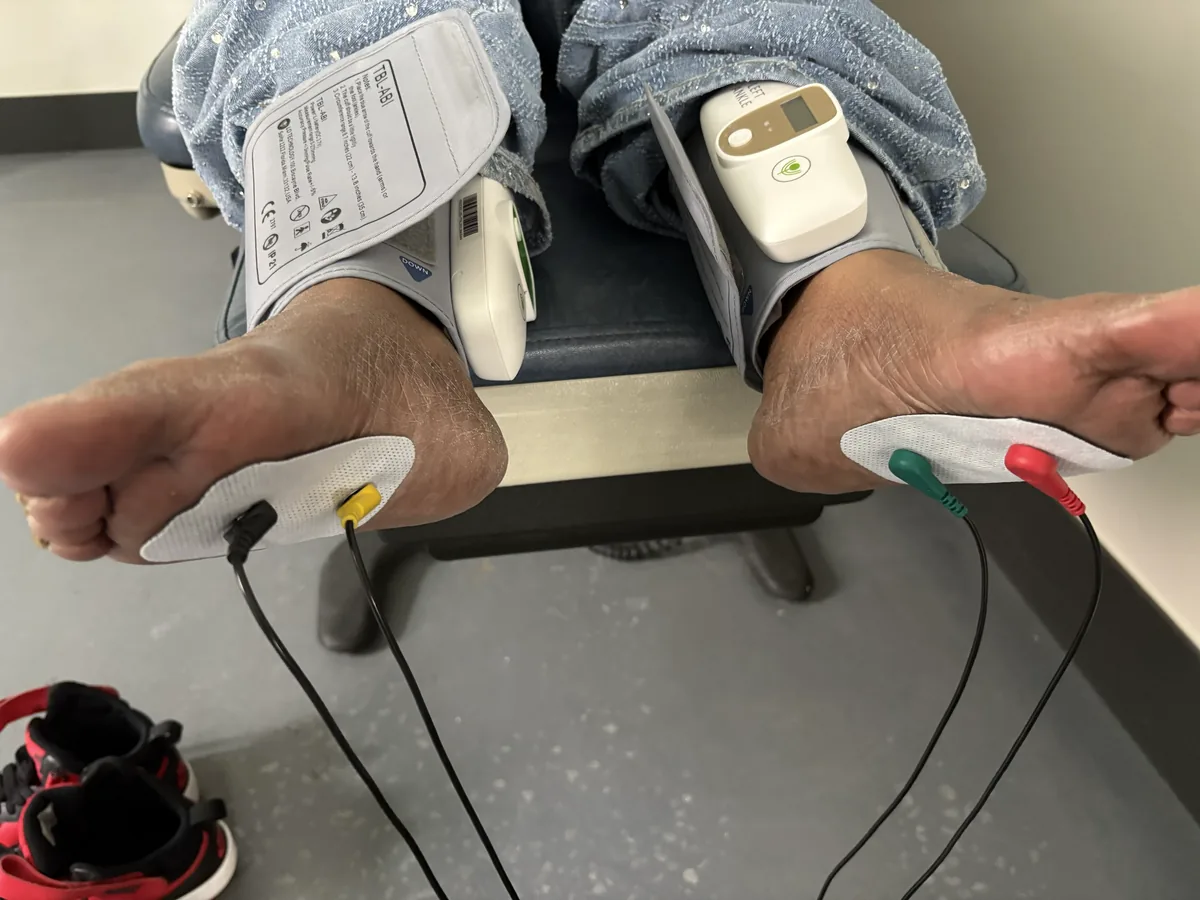
    Patient seated with ankle cuffs and foot electrodes in place for combined vascular and neuropathy testing at Regenerve, 4477 Woodson Rd #104, St. Louis, MO 63134

    The comparison, side by side

    This is a guide to what to ask and what to test, not a self-diagnosis tool. Your clinician makes the diagnosis.

    Distribution

    Length-dependent neuropathy begins distally and climbs [3]. CIDP involves nerve roots as well as peripheral nerve, and presents with proximal as well as distal weakness [1][2].

    Sensory profile

    CIDP emphasizes position and vibration sense over pain and temperature [1]. Length-dependent disease more often opens with small-fiber symptoms, meaning burning and other painful dysesthesias [3].

    Timeline

    CIDP requires persistence of at least eight weeks [1][2]. Length-dependent neuropathy accumulates more gradually and tracks the driver behind it — metabolic, nutritional, toxic, autoimmune or structural.

    How common each is

    CIDP incidence is estimated at 0.15 to 1.6 cases per 100,000 person-years and prevalence at 0.67 to 10.3 per 100,000, with onset typically between 40 and 60 years and a male-to-female ratio of about 2:1 [2]. Distal symmetric polyneuropathy in diabetes is far more common: it affects at least 20% of people with type 1 diabetes after 20 years, and ranges from 10% to 15% of newly diagnosed patients with type 2 diabetes to 50% after 10 years [3].

    When to raise the CIDP question

    Certain features should prompt the question rather than settle it.

    • Weakness that is not confined to the feet, including proximal weakness [1][2]
    • Symptoms that have persisted and evolved beyond eight weeks [1]
    • Sensory loss that emphasizes vibration and position sense over pain and temperature [1]
    • A course that includes relapses rather than steady distal progression [1]

    If your symptoms are changing rapidly, if weakness is severe, or if breathing is affected, that is an emergency evaluation, not a scheduled one.

    How the evaluation is sequenced here

    We perform electrodiagnostic testing (EMG and nerve conduction studies) on site and read it alongside your symptom distribution, examination findings and likely drivers. Because CIDP is uncommon and length-dependent causes are not, the order of the workup follows your pattern and timeline rather than a fixed checklist.

    Where the pattern points to a metabolic, nutritional, toxic or structural driver, testing and diagnosis drive the plan rather than the reverse. That sequence is set out in the Regenerve Protocol for peripheral neuropathy.

    What we do not do

    We do not treat labels, and we do not promise nerve regeneration or reversal of established nerve damage. If the pattern suggests an immune-mediated process such as CIDP, that belongs with the clinicians who manage immune therapy, and our role is to help get the question asked.

    Frequently asked questions

    How long do symptoms have to last before CIDP is considered?

    At least eight weeks. CIDP symptoms must persist for at least 8 weeks to establish the diagnosis [1], and the typical presentation is symmetric progressive proximal and distal muscle weakness with distal numbness evolving over that period [2]. Burning that comes and goes over days is a different question — see small fiber neuropathy symptoms and testing.

    Does burning in my feet point toward CIDP?

    Usually the opposite. CIDP particularly affects position and vibration sense more than pain and temperature [1], while burning and other unpleasant burning sensations are the early small-fiber symptoms of distal symmetric polyneuropathy [3]. What follows from that distinction is covered in peripheral neuropathy treatments for the feet.

    Is CIDP common?

    No. Incidence is estimated at 0.15 to 1.6 cases per 100,000 person-years and prevalence at 0.67 to 10.3 per 100,000 people, with onset typically between ages 40 and 60 and males affected about twice as often as females [2]. That rarity is why the more common drivers are usually worked through first — our sequence is described in our neuropathy services.

    Should I bring my medication list to the evaluation?

    Yes, and the full list including anything started or stopped in the last few years. Medication history is one of the categories we review alongside metabolic, nutritional, toxic and autoimmune drivers. Our discussion of the medications patients ask about most is at statins, metformin and nerve health.

    Start with the Nerve Damage Score

    The Nerve Damage Score is a free five-question assessment that helps identify which driver is most likely behind your symptoms, so your evaluation starts with a direction instead of a guess.

    Regenerve, 4477 Woodson Rd #104, St. Louis, MO 63134. Serving the St. Louis region, Missouri and Illinois, minutes from St. Louis Lambert International Airport.

    Sources

    1. Gogia B, Rocha Cabrero F, Khan Suheb MZ, Lui F, Rai PK. “Chronic Inflammatory Demyelinating Polyradiculoneuropathy.” In: StatPearls [Internet]. Treasure Island (FL): StatPearls Publishing. Last updated 4 March 2024. https://www.ncbi.nlm.nih.gov/books/NBK563249/ — cited for the requirement that symptoms persist at least 8 weeks to establish the diagnosis; inflammation of nerve roots and peripheral nerve; segmental demyelination and remyelination producing onion bulbs; the monophasic, relapsing and progressive courses with about one-third relapsing-remitting; and involvement of position and vibration sense more than pain and temperature.
    2. “Diagnosis of Chronic Inflammatory Demyelinating Polyneuropathy.” Practical Neurology, April 2024 issue. https://practicalneurology.com/diseases-diagnoses/neuromuscular/diagnosis-of-chronic-inflammatory-demyelinating-polyneuropathy/32112/ — cited for the typical presentation of symmetric progressive proximal and distal muscle weakness with distal numbness evolving over at least 8 weeks; the progressive, monophasic and relapsing-remitting courses; incidence of 0.15 to 1.6 cases per 100,000 person-years; prevalence of 0.67 to 10.3 per 100,000; onset around 40 to 60 years; and the approximately 2:1 male-to-female ratio.
    3. Pop-Busui R, Boulton AJM, Feldman EL, Bril V, Freeman R, Malik RA, Sosenko JM, Ziegler D. “Diabetic Neuropathy: A Position Statement by the American Diabetes Association.” Diabetes Care, 2017;40(1):136–154. DOI: 10.2337/dc16-2042. https://pmc.ncbi.nlm.nih.gov/articles/PMC6977405/ — cited for assessment following the typical distal symmetric polyneuropathy pattern starting distally at the dorsal aspect of the great toe on both sides and moving proximally; small-fiber symptoms of pain and burning dysesthesias described as burning, lancinating, tingling or shooting; large-fiber symptoms of numbness, tingling without pain and loss of protective sensation; and prevalence of at least 20% in people with type 1 diabetes after 20 years and 10–15% of newly diagnosed type 2 patients rising to 50% after 10 years.

  • Caught in the Crossfire: How Lupus and Rheumatoid Arthritis Attack Your Nerves

    Caught in the Crossfire: How Lupus and Rheumatoid Arthritis Attack Your Nerves

    When the immune system is dysregulated, nerves are rarely the intended target — but they are frequently the casualties. Autoimmune diseases such as lupus, rheumatoid arthritis, and Sjögren’s syndrome can produce peripheral neuropathy as collateral damage from a body-wide inflammatory process. This article explains the two main routes by which autoimmunity injures nerves, why the resulting neuropathy can take several distinct forms, and why effective treatment must address both the systemic disease and the nerve.

    Nerves as collateral damage

    In an autoimmune disease, the immune system loses the ability to distinguish self from non-self and begins attacking the body’s own tissues. In lupus (systemic lupus erythematosus) and rheumatoid arthritis, the headline targets are joints, skin, kidneys, and other organs — but the same inflammatory machinery can reach the peripheral nerves. Understanding neuropathy in this setting means understanding that the nerve damage is a downstream consequence of a systemic process, which is why simply treating the nerve is not enough.

    Route one: vasculitis — choking the nerve’s blood supply

    Peripheral nerves depend on a network of tiny blood vessels, the vasa nervorum, for their oxygen and nutrients. In autoimmune disease, these vessels can become inflamed — a process called vasculitis. Inflammation thickens and occludes the vessel walls, cutting off blood flow and starving segments of nerve of oxygen (ischemia). The nerve tissue downstream is injured or dies.

    Vasculitic neuropathy often produces a distinctive pattern called mononeuritis multiplex, in which several individual nerves are damaged in a patchy, asymmetric fashion — for example, a wrist drop on one side and a foot drop on the other, rather than the symmetric stocking-glove pattern typical of diabetic neuropathy. This asymmetry is an important clue that points toward a vasculitic, autoimmune cause. Vasculitic neuropathy can progress quickly and is considered a situation that warrants prompt evaluation.

    Route two: direct antibody attack

    The second route is more direct. The autoantibodies and immune complexes generated in autoimmune disease can bind to and attack nerve components themselves, including the myelin sheath and the nerve fibers. Sjögren’s syndrome is particularly associated with sensory neuropathies — including a sensory ganglionopathy in which the sensory nerve cell bodies themselves are targeted — which can cause numbness, imbalance, and painful burning sensations. Different autoimmune conditions favor different antibody targets, which is part of why autoimmune neuropathy is not a single entity but a family of presentations.

    Recognizing the pattern

    Several features raise suspicion that a neuropathy is autoimmune in origin: an asymmetric or patchy distribution (mononeuritis multiplex), a relatively rapid onset, the presence of systemic symptoms (joint pain, rash, dry eyes and mouth, fatigue), and a known or suspected autoimmune diagnosis. When these are present, the workup expands to include autoimmune serologies and, in some cases, nerve conduction studies or nerve biopsy to characterize the process.

    Why treatment must be two-pronged

    This is the central clinical message. Because the neuropathy is driven by a systemic autoimmune process, controlling that process is essential — typically the domain of rheumatologic care, using immune-modulating treatment to quiet the underlying disease. But calming the systemic inflammation does not automatically repair nerves that have already been injured. The nerve itself also needs support: pain management, protection of remaining function, and attention to the same terrain factors — circulation, nutrients, mitochondrial energy — that any injured nerve requires to recover.

    A terrain-focused clinic works alongside a patient’s rheumatologist rather than in place of them: the rheumatologist targets the fire, and the nerve-focused care supports the tissue caught in it. Neither alone is sufficient.

    The overlap with other drivers

    Autoimmune disease also raises the stakes on other neuropathy drivers. Systemic inflammation, certain immune-modulating medications, and reduced activity can all compound nutritional and metabolic contributors, so a complete evaluation still checks the full range of causes rather than stopping at the autoimmune label.

    Frequently asked questions

    How is autoimmune neuropathy different from diabetic neuropathy?

    Diabetic neuropathy is usually symmetric and starts in the feet. Autoimmune neuropathy is often asymmetric and patchy (mononeuritis multiplex) and tends to accompany other systemic symptoms.

    If I control my lupus or RA, will my neuropathy go away?

    Controlling the underlying disease is essential to stop ongoing damage, but nerves already injured need separate support and recover slowly. The two goals go together.

    Can neuropathy be the first sign of an autoimmune disease?

    Sometimes. An unexplained, especially asymmetric, neuropathy can prompt discovery of an underlying autoimmune condition, which is why the workup includes autoimmune testing.

    Should I stop my immune medications if I develop neuropathy?

    No — never adjust these on your own. Some are treating the very process damaging your nerves. Any change is made with your rheumatologist and physician.

    Key takeaways

    • Lupus, RA, and Sjögren’s can damage nerves as collateral damage from systemic autoimmunity.
    • Vasculitis starves nerves of blood, often causing asymmetric mononeuritis multiplex.
    • Direct antibody attack targets myelin and nerve fibers; Sjögren’s favors sensory neuropathies.
    • Asymmetry, rapid onset, and systemic symptoms are clues to an autoimmune cause.
    • Treatment must control the systemic disease and support the injured nerve.

    This article is educational and is not a substitute for evaluation, diagnosis, or treatment by a physician. Individual results vary. Do not start, stop, or change any medication without consulting your physician. Take the free Nerve Damage Score or call/text (314) 886-5902.

    References

    1. Collins MP, Hadden RD. The nonsystemic vasculitic neuropathies. Nat Rev Neurol. 2017;13:302–316.
    2. Mori K, et al. The wide spectrum of clinical manifestations in Sjögren’s syndrome–associated neuropathy. Brain. 2005;128:2518–2534.
    3. Oomatia A, et al. Peripheral neuropathies in systemic lupus erythematosus. Arthritis Rheumatol. 2014.
    4. Gwathmey KG, et al. Vasculitic neuropathies. Lancet Neurol. 2014;13:67–82.

    Find out what is driving your nerve pain

    The free, five-question Nerve Damage Score takes about two minutes and tells you which terrain failure is most likely behind your symptoms.

    Get My Free Nerve Damage Score

    Or call or text (314) 886-5902.

  • Shingles Pain That Won’t Stop? Understanding Postherpetic Neuralgia

    Shingles Pain That Won’t Stop? Understanding Postherpetic Neuralgia

    For most people, shingles is a miserable few weeks and then it’s over. But for a significant minority, the rash heals and the pain doesn’t — it settles into a persistent, often severe nerve pain that can last months or years. This is postherpetic neuralgia (PHN), one of the most common and most distressing forms of neuropathic pain. This article explains why a childhood chickenpox virus can come back decades later to injure nerves, what makes the pain persist, how it is treated, and how it can be prevented.

    A virus that never left

    Shingles is caused by the varicella-zoster virus (VZV) — the same virus that causes chickenpox. After a childhood chickenpox infection, the virus is not eliminated. It retreats into the dorsal root ganglia, the clusters of sensory nerve cell bodies that sit alongside the spinal cord, and lies dormant there, sometimes for decades, held in check by the immune system.

    When immunity wanes — with age, illness, stress, or immune-suppressing conditions and treatments — the virus can reactivate. It travels back down the sensory nerve to the skin, producing the characteristic painful, blistering rash of shingles in a band along the territory of that nerve. But the damage isn’t only skin-deep: the reactivation inflames and injures the nerve and its ganglion along the way.

    Why the pain persists: postherpetic neuralgia

    In postherpetic neuralgia, the nerve injury from the shingles episode leaves the affected sensory pathway damaged and dysregulated. The injured nerves become hyperexcitable and misfire, and the pain-processing system itself can become sensitized, so that even light touch on the healed skin triggers severe pain (a phenomenon called allodynia). The result is persistent burning, stabbing, or electric pain in the area where the rash was, often accompanied by exquisite sensitivity.

    Several factors raise the risk of PHN, most notably older age — the risk climbs substantially with each decade — along with greater severity of the initial shingles episode. This is why prevention and early treatment matter so much.

    Treating postherpetic neuralgia

    PHN is challenging, but there are real tools, and they work best in combination and tailored to the individual.

    Early antiviral treatment of the acute shingles episode (started promptly, within the first days of the rash) can reduce the severity and duration of the outbreak and may lower the risk of persistent pain — one reason to seek care quickly when shingles appears.

    For established PHN, treatments include topical therapies — notably the high-concentration 8% capsaicin patch, which is FDA-approved for postherpetic neuralgia and works by quieting the overactive pain fibers in the affected skin (discussed in detail in the capsaicin article), and lidocaine patches — as well as oral neuropathic-pain medications such as gabapentinoids and certain antidepressants. Because PHN is often localized, the topical, non-systemic options are especially valuable for avoiding whole-body side effects.

    Beyond symptom control, a repair-and-support philosophy addresses the injured nerve terrain itself. The video notes the use of measures aimed at supporting recovery, including cannabinoid-based therapies and glutathione. In the interest of accuracy: cannabinoids have a growing but still-evolving evidence base for neuropathic pain, and glutathione support for nerve recovery is biologically rational but not a proven cure — both belong in the category of adjuncts used within an individualized, physician-guided plan rather than established standalone treatments.

    Prevention: the most important tool

    The single most effective way to deal with postherpetic neuralgia is to prevent the shingles episode that causes it. The recombinant shingles vaccine (Shingrix) is highly effective at preventing shingles and, by extension, PHN, and is recommended for older adults and certain immunocompromised individuals. For anyone in an at-risk group who has not been vaccinated, this is a conversation worth having with a physician — preventing the outbreak is far easier than treating the pain it can leave behind.

    Where this fits

    Postherpetic neuralgia is one of the infection-related drivers of neuropathic pain. Unlike the diffuse, length-dependent neuropathies of metabolic disease, it is typically localized to the nerve territory affected by the shingles outbreak — a distinct pattern that helps identify it and shapes the localized, terrain-supportive approach to treatment.

    Frequently asked questions

    How long does postherpetic neuralgia last?

    It varies widely — from months to years. Some cases resolve gradually; others persist. Older age and a severe initial outbreak increase the likelihood of long-lasting pain.

    Can the capsaicin patch help PHN?

    Yes. The 8% capsaicin patch is FDA-approved for postherpetic neuralgia and can reduce the localized nerve pain by quieting overactive pain fibers; treatment can be repeated.

    Does treating shingles early prevent PHN?

    Prompt antiviral treatment of the acute episode can reduce its severity and may lower the risk of persistent pain, which is why fast care for a shingles rash matters.

    Can I prevent shingles altogether?

    Largely, yes. The recombinant shingles vaccine is highly effective and is recommended for older adults and some others — discuss it with your physician.

    Key takeaways

    • Shingles comes from varicella-zoster virus reactivating from the dorsal root ganglia, injuring the nerve.
    • Postherpetic neuralgia is the persistent nerve pain that can follow, driven by nerve damage and sensitization.
    • Risk rises sharply with age and with a severe initial outbreak.
    • Treatment combines topical options (including the FDA-approved 8% capsaicin patch), oral medications, and terrain support; cannabinoids and glutathione are adjuncts, not proven cures.
    • The shingles vaccine is the most effective way to prevent PHN.

    This article is educational and is not a substitute for evaluation, diagnosis, or treatment by a physician. Individual results vary. Do not start, stop, or change any medication without consulting your physician. Take the free Nerve Damage Score or call/text (314) 886-5902.

    References

    1. Johnson RW, Rice ASC. Postherpetic neuralgia. N Engl J Med. 2014;371:1526–1533.
    2. Gershon AA, et al. Varicella zoster virus infection. Nat Rev Dis Primers. 2015.
    3. Backonja M, et al. NGX-4010 (capsaicin 8% patch) for postherpetic neuralgia: randomized studies. Lancet Neurol. 2008.
    4. Dooling KL, et al. Recommendations of the ACIP for use of recombinant zoster vaccine (Shingrix). MMWR. 2018.

    Find out what is driving your nerve pain

    The free, five-question Nerve Damage Score takes about two minutes and tells you which terrain failure is most likely behind your symptoms.

    Get My Free Nerve Damage Score

    Or call or text (314) 886-5902.

  • Sometimes It’s Not Peripheral Neuropathy: Carpal Tunnel, Sciatica, and Double Crush

    Sometimes It’s Not Peripheral Neuropathy: Carpal Tunnel, Sciatica, and Double Crush

    Not every case of numb hands or burning feet is a systemic, metabolic neuropathy. Sometimes the nerve is simply being squeezed — pinched at the wrist, compressed at the spine, or entrapped somewhere along its path. Mechanical compression is a common and often highly treatable cause of nerve symptoms, and distinguishing it from a diffuse neuropathy changes everything about the treatment. This article explains how compression injures nerves, how to recognize the pattern, and why compression and metabolic disease so often team up through a phenomenon called double crush syndrome.

    How compression injures a nerve

    A nerve under sustained mechanical pressure suffers in two main ways. First, compression restricts blood flow to the nerve (ischemia), starving it of oxygen and nutrients. Second, sustained pressure damages the nerve’s insulating myelin at the site of compression (focal demyelination), disrupting the fast, faithful conduction of signals. If the pressure continues, the underlying nerve fibers themselves can be injured. The result is numbness, tingling, pain, and sometimes weakness — but with a crucial difference from metabolic neuropathy: the problem is localized to the compressed nerve, not spread symmetrically across all the longest nerves.

    Common compression syndromes

    Carpal tunnel syndrome is the classic example: the median nerve is compressed as it passes through a tight tunnel at the wrist. It typically causes numbness and tingling in the thumb, index, middle, and part of the ring finger, often worse at night, and sometimes weakness of grip. It is one of the most common nerve disorders and is frequently very treatable.

    Radiculopathy (including sciatica) occurs when a nerve root is compressed as it exits the spine — for example, by a herniated disc or arthritic narrowing. Sciatica is the well-known form: pain, numbness, or weakness radiating from the low back down the leg along the path of the affected nerve root. Because the compression is at the spine, the symptoms follow a specific nerve’s territory rather than a stocking-glove pattern.

    Other entrapments (such as the ulnar nerve at the elbow) follow the same logic: a specific nerve, compressed at a specific spot, producing symptoms in that nerve’s specific distribution.

    Recognizing compression versus diffuse neuropathy

    The pattern is the tell. Metabolic neuropathies (like diabetic neuropathy) are usually symmetric and length-dependent — both feet first, then moving upward. Compression syndromes are usually focal and asymmetric — one nerve, one territory, sometimes provoked by particular positions or activities (typing, a night of a bent wrist, prolonged sitting). Nerve conduction studies and electromyography can localize where along a nerve the problem lies, helping confirm compression and pinpoint its site. Getting this distinction right matters, because a compressed nerve may be relieved by decompression — mechanical or surgical — whereas a metabolic neuropathy needs a metabolic approach.

    Double crush syndrome: when compression and metabolism combine

    Here is one of the most clinically important and underappreciated concepts in nerve medicine. In 1973, Upton and McComas, writing in The Lancet, proposed double crush syndrome: the idea that a nerve compressed at one point becomes more vulnerable to injury at a second point along its length. A single mild compression that might not cause symptoms on its own can become symptomatic when combined with a second insult.

    The insight extends beyond two mechanical compressions. A nerve that is metabolically stressed — by diabetes, by nutritional deficiency, by toxins — is already compromised, and that makes it far more susceptible to symptomatic injury from even modest mechanical compression. This is why so many patients have both: a metabolic neuropathy that has lowered the nerve’s reserve, plus a compression (like carpal tunnel) that pushes it over the threshold into symptoms. Treating only one of the two often leaves the patient frustrated.

    Why a dual approach works best

    The double-crush concept has a direct treatment implication: address both the mechanical and the metabolic contributors. Relieving the compression — through ergonomic changes, splinting, injections, physical therapy, or, when appropriate, decompression procedures — removes the physical insult. Simultaneously supporting the nerve’s metabolic terrain — blood sugar, nutrients, mitochondrial energy, inflammation — raises its resilience so it can tolerate normal life and heal. Neither alone fully solves a double-crush situation; together they can. This dual philosophy is exactly why a thorough evaluation checks for compression even in someone with known metabolic disease, and checks for metabolic drivers even in someone with an obvious entrapment.

    Where this fits

    Mechanical compression is the mechanical category in the three-domain framework of neuropathy drivers — and double crush syndrome is the bridge that explains why the categories so often overlap. A complete workup deliberately looks across all of them, because the most common real-world scenario is not a single cause but a combination, each amplifying the others.

    Frequently asked questions

    How do I know if it’s carpal tunnel or a general neuropathy?

    Carpal tunnel causes symptoms in a specific hand distribution, often worse at night, on one or both sides. Diffuse neuropathy is usually symmetric and starts in the feet. Nerve conduction studies can distinguish and localize the problem.

    Can I have both compression and neuropathy?

    Yes — and it’s common. Double crush syndrome describes how a metabolically stressed nerve becomes more vulnerable to compression, so the two frequently coexist and compound each other.

    Is surgery always needed for compression?

    No. Many compression syndromes respond to conservative measures — splinting, ergonomics, injections, therapy. Decompression is considered when conservative care is insufficient or damage is progressing.

    Why didn’t treating my diabetes fix my hand numbness?

    Because the hand numbness may be a compression (like carpal tunnel), not the diabetic neuropathy — a classic double-crush situation where both the mechanical and metabolic sides need attention.

    Key takeaways

    • Not all nerve pain is metabolic; mechanical compression is common and often treatable.
    • Carpal tunnel and sciatica compress a specific nerve, causing focal, asymmetric symptoms.
    • Compression injures nerves via ischemia and focal demyelination; the pattern distinguishes it from diffuse neuropathy.
    • Double crush syndrome (Upton & McComas, 1973) explains why metabolically stressed nerves are more vulnerable to compression.
    • The best results come from addressing both the mechanical and metabolic contributors together.

    This article is educational and is not a substitute for evaluation, diagnosis, or treatment by a physician. Individual results vary. Take the free Nerve Damage Score or call/text (314) 886-5902.

    References

    1. Upton ARM, McComas AJ. The double crush in nerve-entrapment syndromes. Lancet. 1973;2(7825):359–362.
    2. Padua L, et al. Carpal tunnel syndrome: clinical features, diagnosis, and management. Lancet Neurol. 2016;15:1273–1284.
    3. Wilbourn AJ, Gilliatt RW. Double-crush syndrome: a critical analysis. Neurology. 1997;49:21–29.
    4. Rempel D, et al. Pathophysiology of nerve compression syndromes. J Bone Joint Surg Am. 1999.

    Find out what is driving your nerve pain

    The free, five-question Nerve Damage Score takes about two minutes and tells you which terrain failure is most likely behind your symptoms.

    Get My Free Nerve Damage Score

    Or call or text (314) 886-5902.